Whole Body Imaging - I 131
View main image(tr) in a separate image viewer
Full history/Diagnosis is available below
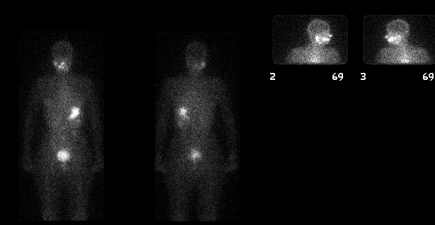
(2) No evidence of new metastatic spread. No activity within the lungs is appreciated. Physiologic distribution of tracer activity is appreciated in the salivary glands; stomach; bladder; and, kidneys. Asymmetric salivary gland activity is seen, a reasonably common finding.
(2) The average dose used for diagnostic imaging at our institution is 5 mCi in adults The oral I-131 is swallowed on the first day of treatment; and delayed images are obtained 48 hours later. A large field of view camera calibrated for 364 kev with a 20% window and a high energy parallel hole collimator is used. Generally, images are collected for 100k or 10 minutes, whichever occurs first. Ocassionally, the patient may be imaged after an ablative dose of I-131 (eg, 100 mCi), with imaging typically 3-5 days after the treatment dose. Increased count rate may change the time of acquisition in the post treatment setting.
(3) I-123 can be used as an alternate imaging agent.
(2) The patient's TSH should be adequately elevated (>20 microunits/ml) to insure good uptake by residual thyroid tissue and/or metastatic foci. This is typically done by withdrawal of thyroid hormone for 2-3 weeks, but can be done with administration of TSH (Thyrogen). Thyrogen is typically used only when prior withdrawal scans have been negative.
(3) The patient should have avoided iodine-containing mediciations (such as amiodarone, SSKI, IV contrast, Lugol's solution or other iodine-containing products) during 1-2 months prior to the examination. The effect of amiodarone can last 1-2 years.
(4) A nonpregnant state of all females within child-bearing age should be confirmed prior to I-131 dosing.
References and General Discussion of Thyroid Scintigraphy (Anatomic field:Face, Mastoids, and Neck, Category:Neoplasm, Neoplastic-like condition)
Return to the Teaching File home page.