Case Author(s): Akash Sharma, M.D. and Henry D. Royal, M.D. , September 20, 2004 . Rating: #D3, #Q5
Diagnosis: Post-traumatic splenosis
Brief history:
46-year-old woman with an abdominal mass incidentally found on a recent outside renal stone CT study.
Images:

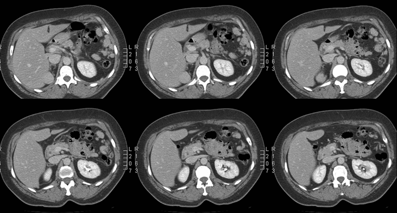
Selected images from initial abdomen CT
View main image(ct) in a separate image viewer
View second image(si).
Planar images
View third image(si).
Selected axial images (SPECT)
Full history/Diagnosis is available below
Diagnosis: Post-traumatic splenosis
Full history:
A 46-year-old patient presents with history of abdominal injury and subsequent splenectomy at 14 years of age. On a recent outside renal stone CT protocol study, a mass was incidentally found adjacent to the superior pole of the right kidney. Additionally, multiple nodular masses were seen in the left upper quadrant. While the left upper quadrant masses were thought to be from splenosis, the patient presents to us for evaluation for the right perinephric mass.
Radiopharmaceutical:
1.97 mCi Tc-99m heat-damaged in-vitro-labeled autologous red blood cells i.v.
Findings:
Abdomen and Pelvis CT (done first): Mass in the right upper quadrant appears to be separate from the liver, right adrenal, and kidney. The patient has multiple similar masses in the left upper quadrant which appear to be splenules and it may be that she has had a previous splenic trauma and the right upper quadrant mass is also a splenule. Alternatively, if the patient has a history of lymphoma, this could be an unusual manifestation of adenopathy. A heat damaged tag red cell scan is recommended for further evaluation. Kidney stones identified in the left kidney
Splenic imaging: Images of the abdomen show multiple foci of radiotracer activity in the left upper quadrant, adjacent to the superior pole of the right kidney, adjacent to the inferior tip of the right lobe of the liver, and two smaller faint foci in the mid abdomen region
All of these lesions correlate with multiple homogeneously enhancing nodular masses seen on the CT performed at Mallinckrodt Institute of Radiology.Additional planar images of the thorax were obtained which demonstrate no increased radiotracer activity in the thorax.
Discussion:
In the setting of suspected splenosis as a result of prior trauma, imaging features play a key role in the diagnosis of ectopic splenic tissue, especially since the differential diagnosis would include more worrisome entities such as malignancies, especially lymphoma.
Splenosis itself may induce relapse of hematologic diseases, mainly autoimmune thrombocytopenia. Given that multiple presumed splenules were identified in the left upper quadrant in this patient, a heat damaged red blood scan is the ideal study to determine the nature of the right upper quadrant lesion.
Since the splenules in the left upper quadrant will expectedly take up the heat-damaged red blood cells, an internal comparison was available to evaluate the nature of right upper quadrant mass. A confirmatory imaging pattern will eliminate the need for any invasive tests such as a needle biopsy.
Hagman TF, Winer-Muram HT, Meyer CA, Jennings SG. Intrathoracic splenosis: superiority of technetium Tc 99m heat-damaged RBC imaging.
Chest. 2001 Dec;120(6):2097-8.
View followup image(mc).
Comparison of selected contrast-enhanced abdomen CT images and heat damaged RBC SPECT study.
Differential Diagnosis List
Based on CT findings: splenosis vs. lymphoma vs. mets.
Based on combined CT and NM findings: Splenosis.
ACR Codes and Keywords:
References and General Discussion of Spleen Imaging (Anatomic field:Vascular and Lymphatic Systems, Category:Effect of Trauma)
Search for similar cases.
Edit this case
Add comments about this case
Return to the Teaching File home page.
Case number: si005
Copyright by Wash U MO

