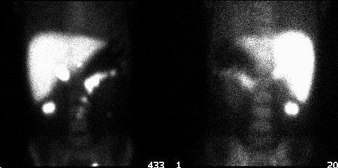
Anterior and posterior images are shown.
View main image(si) in a separate image viewer
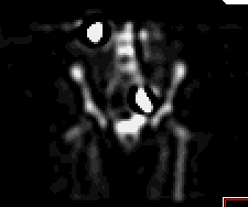
View second image(si). Coronal SPECT image.
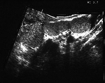
View third image(us). Pelvic ultrasound
Full history/Diagnosis is available below
2. The heat-damaged red cell study reveals several foci of high uptake in the left pelvis (correlating with the nodular lesions seen on ultrasound), left upper quadrant and right upper quadrant. These findings are consistent with splenosis.
Splenosis results from autotransplantation of splenic tissue after trauma. It occurs in up to two-thirds of patients with splenic rupture. Although most patients remain asymptomatic, some will experience pain or develop a palpable mass that may be mistaken for a neoplasm. In rare cases complications may develop including bleeding due to deep invasion of splenic tissue into tissue, torsion or obstruction of bowel. Splenic implants are frequently numerous, draw their blood supply from the tissue to which they bind, and do not have a well- defined capsule. Splenosis is distinct from accessory splenic tissue. Accessory spleens (which have an incidence of 10-40% of patients at autopsy) are generally few in number, commonly occur at the spleno-pancreatic ligament or other structures supplied by the splenic artery, and have a normal hilum and capsule. The incidence of splenosis after splenectomy for trauma is high. In one series (Gunes, et al., 1994), the incidence was 58% and the most sensitive technique for detecting the splenosis was selective splenic scintigraphy with SPECT, utilizing heat-denatured red blood cells (as opposed to planar imaging alone or imaging with liver-spleen scintigraphy utilizing Tc-99m sulfur colloid).
Twenty-five percent of the body lymphoid tissue is located in the spleen. One function of the spleen is to remove particulate antigens. The incidence of sepsis due to encapsulated bacteria increases after a splenectomy. This incidence appears to be slightly lower in patients who have had a splenectomy due to trauma, possibly due to subsequent development of splenosis and some restoration of the immunologic function of the spleen. Approximately 0.1% of splenectomized patients will develop a fatal sepsis (usually within 2 years of splenectomy).
The spleen may be imaged scintigraphically with the liver-spleen agent, Tc-99m sulfur colloid. Approximately 10% of the radiopharmaceutical will be taken up by the spleen in normal patients. Imaging of the spleen specifically may be performed with Tc-99m heat-denatured red blood cells. Approximately 90% of the radiopharmaceutical will be taken up by spleen. Heat-denatured red blood cells are better in post-splenectomy cases in which small foci are expected and in which foci may be found adjacent to the liver.
The preparation of heat-damaged red blood cells begins with pre-tinning red blood cells in vivo by injecting stannous pyrophosphate. After waiting 20 minutes, collect 5 ml of blood into a heparinized syringe. Wash twice with 20 ml saline. Heat 2 ml of the packed RBC’s in a glass container at 49.5° C for 20 minutes. Wash with 20 ml saline. Label with pertechnetate. If heated for too short a time or at too low a temperature, there will be significant blood pool activity (it will appear similar to a regular tagged RBC study). If heated too long or at too high a temperature, there will be more general reticuloendothelial uptake, with a higher degree of liver uptake.
References:
Gunes I, Yilmazlar T, Sarikaya I, Akbunar T and Irgil C. Scintigraphic detection of splenosis: superiority of tomographic selective spleen scintigraphy. Clin Radiol. 49: 115-117, 1994.
Livingston CD, Levine BA, Lecklitner ML and Sirinek KR. Incidence and function of residual splenic tissue following splenectomy for trauma in adults. Arch Surg. 118: 617-620, 1983.
Solheim K and Nerdrum HJ. Scintigraphic follow-up of splenic rupture. Clin Nuc Med. 10: 851-854, 1985.
References and General Discussion of Spleen Imaging (Anatomic field:Gasterointestinal System, Category:Normal, Technique, Congenital Anomaly)
Return to the Teaching File home page.