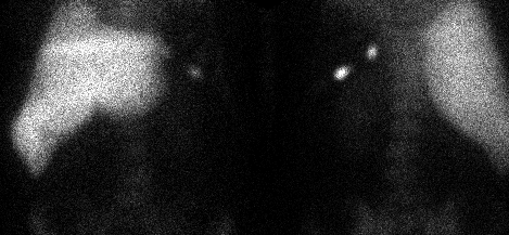
Anterior and posterior images of the chest and abdomen obtained 30 minutes after injection of Tc-99m in vitro-labeled heat-damaged red blood cells.
View main image(si) in a separate image viewer
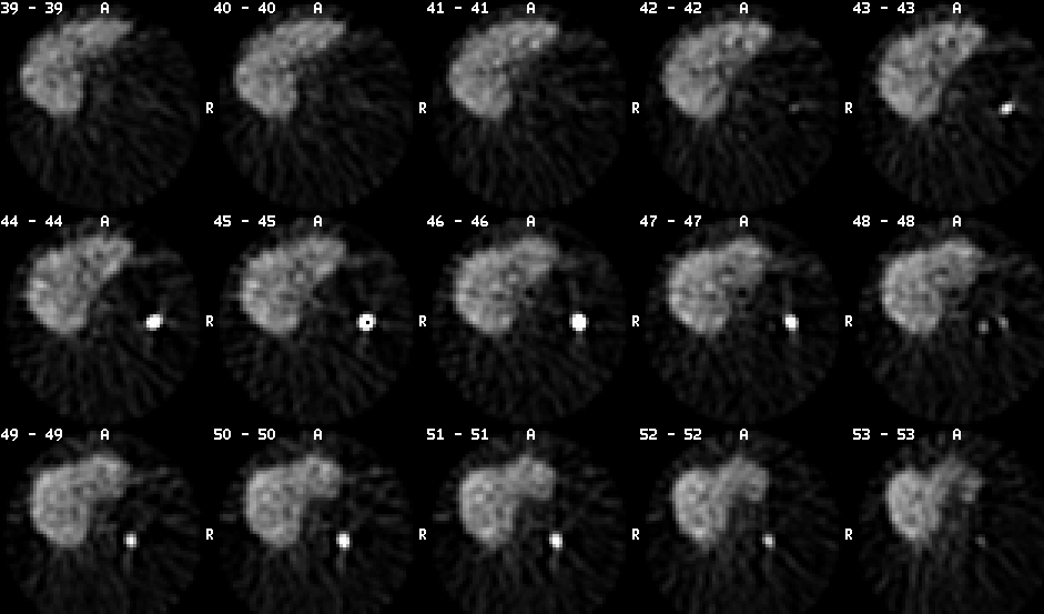
View second image(si). Axial SPECT images performed 40 minutes after injection of Tc-99m in vitro-labeled heat-damaged red blood cells.
Full history/Diagnosis is available below
Following splenectomy, significant improvement occurs in 70-90% of patients and permanent, complete remission occurs in 45-60% of patients . Even if splenectomy does not produce adequate improvement in the more severe cases, the absence of the splenic tissue usually facilitates management by decreasing the steroid requirement. Susceptibility to infection is increased following splenectomy, but the risk is slight in adults compared with children.
Accessory splenic tissue may be responsible for a failure to respond to splenectomy in the immediate postoperative period or a recurrence of thrombocytopenia several months to years later. In one study, of those patients who underwent accessory splenectomy, all had normalization of platelet counts, but less good results have been obtained in other studies. Because accessory spleens are found in 16-19% of patients at the time of splenectomy, careful search must be made for this tissue. The presence of Howell-Jolly bodies on the peripheral blood smear after splenectomy does not ensure the absence of an accessory spleen.
Imaging with Tc-99m heat-damaged red cells is generally considered to be a better technique for finding small accessory spleens (or deposits of splenosis) than is imaging with Tc-99m sulfur colloid. Accessory splenic tissue has also been detected on images obtained after injection of In-111 labeled platelets.
Reference: Hemostasis and Thrombosis, second edition, editor Robert Colman; Lippincott, 1987.
References and General Discussion of Spleen Imaging (Anatomic field:Vascular and Lymphatic Systems, Category:Organ specific)
Return to the Teaching File home page.