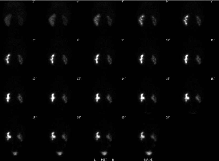

Initial posterior images.
View main image(rs) in a separate image viewer
View avi cine image (1 meg) Posterior images after furosemide administration
View third image(rs). Time/activity curves and analysis
View fourth image(gu). Anterior image of voiding cystourethrogram
Full history/Diagnosis is available below
Renogram after furosemide administration shows prompt clearance of residual pelvicaliceal activity of the left. On the right, there are intermittent episodes of increasing radiotracer activity in the collecting system consistent with marked vesicoureteral reflux. The reflux is most evident on cine images, and was surprisingly difficult to see on static images (not shown). This was due to the fact that the static images were reframed to 2 minute frames for filming purposes (as opposed to the 20-second frames available for cine).
Time/activity curves and analysis demonstrate that the right kidney contributing 28% and the left kidney 72% of total renal function. Also the T1/2 of clearance of residual activity in the left renal collecting system after furosemide administration is 6 minutes, normal, and suggesting no obstruction on the left.
Radiograph taken during a VCUG shows grade V vesicoureteral reflux on the right.
When performing a renogram on an infant or young child, a Foley catheter should be in place to drain urine from the bladder. In this patient the Foley catheter was present but not functioning properly, thus the patient's marked vesicoureteral reflux is well demonstrated.
Viewing post-diuretic images in cine format is helpful to detect reflux.
References and General Discussion of Renal Scintigraphy (Anatomic field:Genitourinary System, Category:Normal, Technique, Congenital Anomaly)
Return to the Teaching File home page.