Case Author(s): Rusty Roberts, M.D. and Tom R. Miller, M.D., Ph.D. , 9/24/05 . Rating: #D2, #Q3
Diagnosis: Urine leak
Brief history:
An 18-year-old man who sustained a right flank gunshot wound which traversed his abdomen and lodged in his left flank one week prior to this examination. He currently has a left lower quadrant Jackson-Pratt drain which has an elevated creatinine of 10.3mg/dL. He has had normal urine output by Foley catheter.
Images:

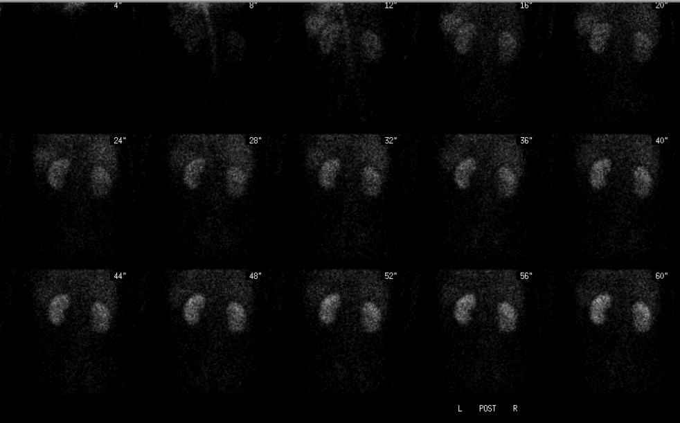
Posterior angiographic phase
View main image(rs) in a separate image viewer
View second image(rs).
Dynamic posterior images through 20 minutes
View third image(rs).
Delayed posterior, anterior and bilateral lateral abdominal images
View fourth image(ct).
Transaxial contrast CT at the renal level
Full history/Diagnosis is available below
Diagnosis: Urine leak
Full history:
This 18-year-old gman was transfered after a gunshot wound one week previously which he sustained to his right posterior lateral subcostal area with a perforation of the inferior lateral border of the right lower lobe of his liver and a right pneumothorax. He also had a laceration of his right kidney as well as the upper pole of his left kidney. The inferior pole of his spleen was also lacerated. The patient had an exploratory laparotomy with an abdominal drain placed before transfer.
The drainage had not subsided after placement of the drain, and a urine leak was suspected. Creatinine was 10.3mg/dL in the drain fluid. The renal scintigraphy was requested to determine function and localize the injury.
Radiopharmaceutical:
7.9 mCi Tc-99m MAG3 i.v.
Findings:
The posterior abdominal radionuclide angiogram demonstrates prompt, symmetrical perfusion of the kidneys. Sequential renal images demonstrate normal prompt uptake within the renal cortex and prompt excretion into intra-renal collecting systems bilaterally.
There is progressive accumulation of activity adjacent to the left kidney extending from the left hemidiaphragm caudally to the mid-abdominal level. This is contained within the left abdomen. The right kidney appears intact without leak. There is radionuclide within the right ureter and bladder as well as the Foley catheter. Subsequent imaging of the Jackson-Pratt drainage bag showed no evidence of radionuclide uptake. There is no definite activity within the left ureter indicating that the majority of urine from the left kidney was likely leaking into the pararenal space.
The split renal function for the right was 43%, and the left was 57%.
Discussion:
Imaging with Tc-99m MAG3 is an effective way to diagnose a renal leak.
Followup:
The patient had a cystoscopy and retrograde pyelogram the same day as the renal scintigraphy, revealing a renal laceration involving the left renal pelvis with intact calyces. A left ureteral stent was placed. Two weeks later the Foley catheter and abdominal drain were removed. The stent was removed six weeks after placement.
Major teaching point(s):
These findings likely representto a urine leak into the para-renal space; however, a leak into the peri-renal space and leak into the peritoneal space cannot be excluded.
Renal function aids in surgical planning, in this case showing good function indicating that a nephrectomy is not indicated, with a stent the preferred option.
Differential Diagnosis List
Urine leak from calyx, renal pelvis, or ureter.
ACR Codes and Keywords:
References and General Discussion of Renal Scintigraphy (Anatomic field:Genitourinary System, Category:Effect of Trauma)
Search for similar cases.
Edit this case
Add comments about this case
Return to the Teaching File home page.
Case number: rs033
Copyright by Wash U MO

