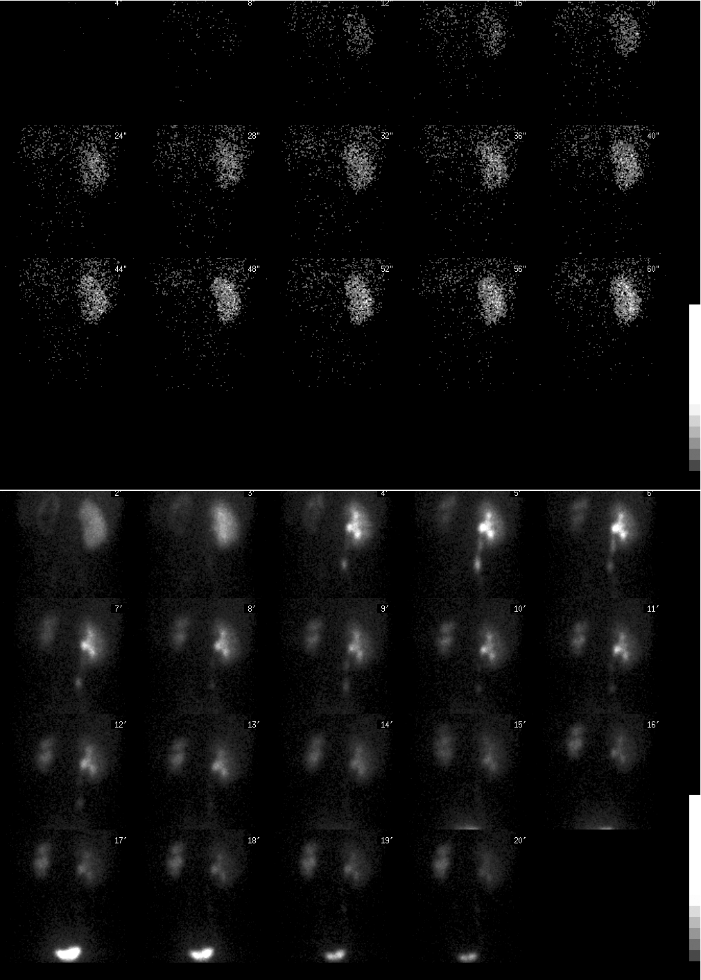

Top panel: Posterior projection images (2 sec/fr for 1 min) after injection of radiopharmaceutical. Bottom panel: Posterior projection images (1 min/fr for next 20 min) after injection of radiopharmaceutical.
View main image(rs) in a separate image viewer
View second image(rs). Renal function curves.
View third image(rs). Post Lasix posterior projection images (1 min/fr for 33 min).
View fourth image(rs). Left Panel: Initial post void image following 20 minutes of imaging Right Panel: Post void image after 33 minutes of imaging following the administration of Lasix.
Full history/Diagnosis is available below
Upper Gastrointestinal series with small bowel follow through demonstrates mild gastoesophageal reflux. The examination was otherwise normal with normal small bowel transit and no evidence for malrotation. Abdominal sonogram demonstrates an enlarged left kidney with severe hydronephrosis and cortical thinning. There was no hydroureter. The right kidney was enlarged (compensatory hypertrophy). The bladder was unremarkable.
Renal scintigraphy was requested to evaluate renal function and severity of obstruction.
RIGHT KIDNEY: On initial radionuclide angiogram, there is normal uptake of radiopharmaceutical activity. The kidney is slightly enlarged but normal in contour. There is normal accumulation and excretion of radiopharmaceutical in the collecting system. The right ureter and bladder are normal
FUNCTION CURVE: Right kidney, normal functional curve, 89% of total renal function. Left kidney, decreased renal function, 11% of total renal function.
LEFT KIDNEY, POST LASIX: There is slow clearance of radiopharmaceutical from a dilated collecting system. The half time of tracer clearance from the left kidney and collecting system is 41 minutes (see follow up image). This is consistent with obstruction of the collecting system.
RIGHT KIDNEY, POST LASIX: There is prompt excretion of activity in the right collecting system. There is no evidence for obstruction of the right collecting system.
Contrast intravenous urography and sonography cannot reliably distinguish obstructive from nonobstructive causes of hydronephrosis.
The evaluation of function in the presence of obstruction does not provide a reliable indication for potential recovery of function following surgery. High pressure in the collecting system result in reduction in renal blood flow and function.
The most common cause of unilateral obstruction is from obstruction at the ureteropelvic junction (UPJ). This can be due to an intrinsic ureteral narrowing of an aperistaltic segment or extrinsic compression from an aberrant crossing renal vessel. Severe vesicoreteral reflux can result in ureteral elongation, kinking and eventual mechanical obstruction at the UPJ.
REFERENCES: Mandell GA, et al. "Society of Nuclear Medicine Procedure Guideline for Diuretic Renography in Children." Society of Nuclear Medicine Guidelines Manual, June 2002. 159-63.
Follow up renal scintigraphy 5 months later demonstrates some interval improvement in flow and function in the left kidney. The left kidney now contributes 19% of total renal function. Following administration of lasix, there is prompt clearance of activity in the left kidney with a half time clearance of 2 minutes, normal.
View followup image(rs). Renal scintigraphy follow up 5 months after initial renal scan.
Upper Left panel: Posterior projection images (1 min/fr x 20 min) excluding first minute
Upper Right panel: Posterior projection images post lasix (1 min/fr x 20 minutes)
Bottom Left panel: Renal function curves
Bottom Right panel: Post lasix washout curves of the left kidney before and after pyleoplasty
References and General Discussion of Renal Scintigraphy (Anatomic field:Genitourinary System, Category:Organ specific)
Return to the Teaching File home page.