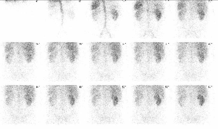
Blood-flow images in the posterior projection
View main image(rs) in a separate image viewer
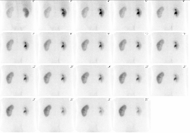
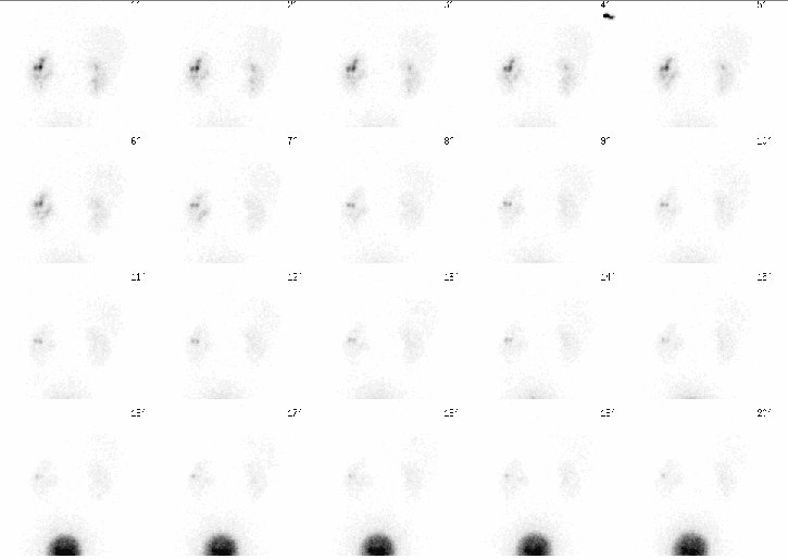
View second image(rs). Twenty-minute dynamic images
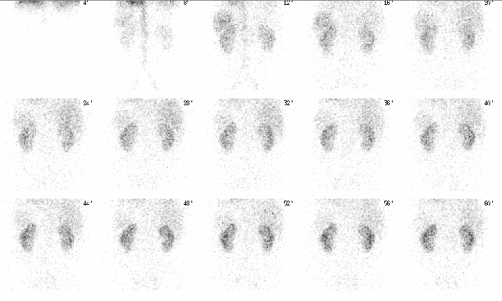
View third image(rs). Blood-flow images performed one month later
View fourth image(rs). Dynamic images performed one month later. Note: The patient has a stent in the left collecting system.
Full history/Diagnosis is available below
A spiral CT scan from an outside hospital (not available) demonstrated left hydronephrosis without evidence of obstructing calculi, suggesting UPJ obstruction.
Following successful stenting of the left collecting system, repeat renal scintigraphy demonstrated marked improvement in function of the left kidney.
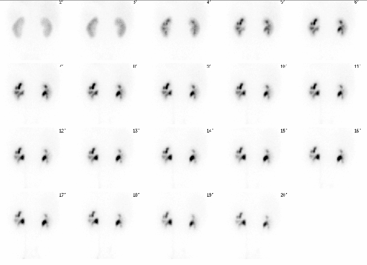
Furosemide was administered (shown below) to evaluate for high-flow obstruction with the stent in place. There was no evidence of obstruction.
Diruetic renal scintigraphy is useful for evaluation of high-flow states that can cause obstruction.
View followup image(rs). Dynamic post-furosemide images performed one month after the initial study are shown. Note: Left collecting system stent in place.
A Foley catheter is sometimes useful to minimize downstream pressure in patients who cannot void well and in patients with reflux as well as to prevent diaper or urine contamination during the long imaging sequence.
References and General Discussion of Renal Scintigraphy (Anatomic field:Genitourinary System, Category:Organ specific)
Return to the Teaching File home page.