Case Author(s): Edward Pinkus,M.D. and Jerold Wallis,M.D. , 3/21/02 . Rating: #D3, #Q4
Diagnosis: Hypoperfusion of kidney secondary to dissection of the aorta.
Brief history:
47 year old man with type B aortic dissection.
Images:

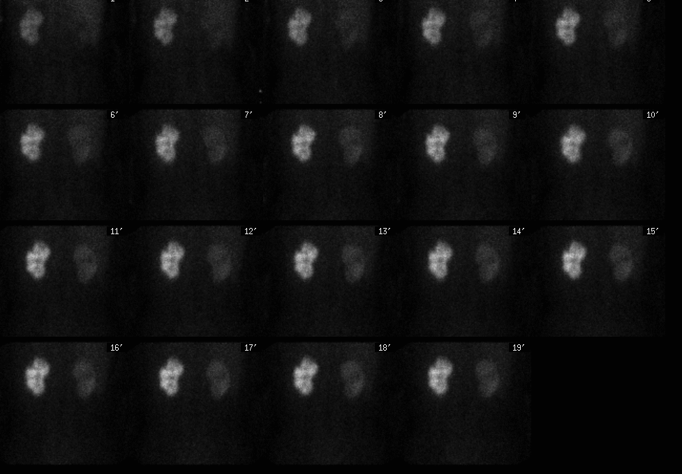
Posterior renal images
View main image(rs) in a separate image viewer
View second image(rs).
Posterior renal images, now status post right renal artery stent placement.
View third image(ct).
Mid-sagittal reconstructed CT image of chest and abdomen.
Full history/Diagnosis is available below
Diagnosis: Hypoperfusion of kidney secondary to dissection of the aorta.
Full history:
47 year old man with an acute type B aortic dissection, status post stenting of the supraceliac aorta, bilateral common iliac arteries, and left renal artery, presents with acute elevation of creatinine and was found to have markedly decreased perfusion of the right kidney by renal scintigraphy. Patient underwent stent placement of the right renal artery and follow-up renal scintigraphy shows improved perfusion and function of the right kidney.
Radiopharmaceutical:
7.8 mci and 8.0 mci of Tc-99m MAG-3 i.v.
Findings:
Initial renal scintigraphy:
Markedly decreased perfusion and function of right kidney, multiple wedge shaped perfusion defects of the left kidney, which correlate to areas of infarction seen on prior CT. Progressive accumulation in the parenchyma, which suggestive of (but not specific for) acute tubular necrosis.
Post stent placement renal scintigraphy:
Markedly improved perfusion and function of the right kidney and progressive accumulation in the parenchyma of both kidneys, suggestive of (but not specific for) acute tubular necrosis.
CT-sagittal reconstruction:
Type B aortic dissection.
Discussion:
Renal perfusion abnormalities may be encountered in patients evaluated for unexplained renal failure. Renal artery occlusion, stenosis, venous thrombosis, and renal infarction can be demonstrated by nuclear technique.
ACR Codes and Keywords:
References and General Discussion of Renal Scintigraphy (Anatomic field:Genitourinary System, Category:Organ specific)
Search for similar cases.
Edit this case
Add comments about this case
Return to the Teaching File home page.
Case number: rs028
Copyright by Wash U MO

