Case Author(s): Mark Fister, M.D. and Tom R. Miller, M.D., Ph.D. , 05/17/01 . Rating: #D2, #Q4
Diagnosis: Post-transplant urinoma
Brief history:
A 52 year-old man who underwent cadaveric renal transplant one week ago. He now presents with decreased urine output.
Images:

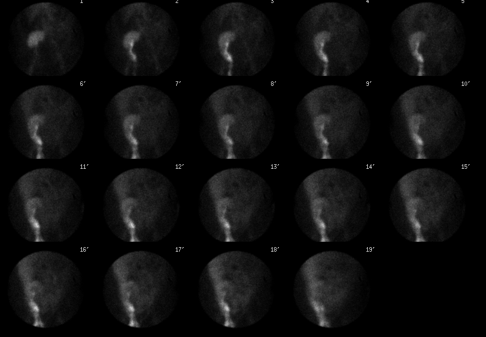
Dynamic Anterior Images, 1 minute frames.
View main image(rs) in a separate image viewer
View second image(rs).
Static Images of the scrotum and urinary catheter bag.
Full history/Diagnosis is available below
Diagnosis: Post-transplant urinoma
Full history:
A 52 year-old man who underwent cadaveric renal transplant one week ago now presents with decreased urine output. Physical examination reveals massive scrotal enlargement beginning with the onset of oliguria. A renal ultrasound revealed new, marked ascites throughout the abdomen and pelvis.
Radiopharmaceutical:
Tc99m-MAG3, i.v.
Findings:
Tc99m-MAG3 images demonstrate prompt uptake and excretion of the radiopharmaceutical. Tracer quickly flows into the scrotum and diffuses throughout the peritoneal space where it accumulates over time. An image of the Foley bag confirms the lack of significant urine output.
Discussion:
Urinomas, though usually found between the transplanted kidney and the bladder, can occur in unexpected locations such as the scrotum or thigh, and can even rupture intraperitoneally to produce urine ascites. Urinomas result from continued slow leakage of urine from any portion of the urinary collecting system, though they typically originate from the ureteroneocystostomy. Ureteral breakdown may result from vascular insufficiency leading to ureteral necrosis. Alternatively, distal obstruction can result in ureteral breakdown.
An abrupt drop in urine output from a previously normally functioning transplant may suggest the development of a urinoma, and swelling may occur around the graft, in the ipsilateral leg, or within the scrotum or labia. Clear fluid draining from the wound is an ominous sign.
Sonographically, urinomas are typically anechoic, non-septated fluid collections that can quickly increase in size. On radionuclide examination, they typically appear as photopenic defects which gradually accumulate tracer. Poor renal excretion may require delayed images.
Followup:
Surgery revealed urine leaking from the superior ridge of the bladder anastomosis with approximately 3 liters of fluid in the abdominal cavity and 600 cc in the wound and scrotum. The ureter was re-anastamosed at another site with subsequent placement of a double-J internal stent.
Major teaching point(s):
Urinomas are a common cause of post-transplant fluid collections that can occasionally extend a significant distance from the kidney. Renal scintigraphy can be extremely helpful for determining their origin.
Reference: Becker JA, Choyke PL, et al. “ Imaging the Transplanted Kidney.” pp. 2668-70 in Clinical Urography. ed. Pollack, HM. W.B. Saunders, Philadelphia 2000.
ACR Codes and Keywords:
References and General Discussion of Renal Scintigraphy (Anatomic field:Genitourinary System, Category:Organ specific)
Search for similar cases.
Edit this case
Add comments about this case
Read comments about this case
Return to the Teaching File home page.
Case number: rs026
Copyright by Wash U MO

