Case Author(s): Gabriel De Simon, M.D. and Tom R. Miller, M.D., Ph.D. , 02/02/2001. . Rating: #D3, #Q4
Diagnosis: Renal transplant urinary leak.
Brief history:
Three weeks after renal transplantation, with diminishing urine output.
Images:

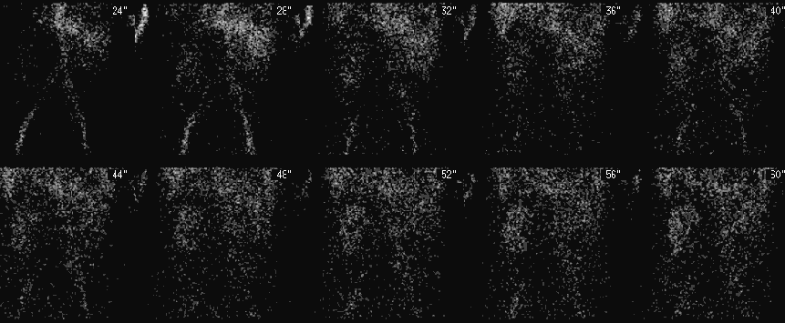
Anterior angiographic flow images of the abdomen and pelvis.
View main image(rs) in a separate image viewer
View second image(rs).
Anterior images of the abdomen and pelvis of the first 20 minutes after injection of the tracer.
View third image(rs).
7-hour delayed anterior image of the abdomen and pelvis.
View fourth image(us).
Longitudinal and transverse ultrasound images of the renal transplant.
Full history/Diagnosis is available below
Diagnosis: Renal transplant urinary leak.
Full history:
Thirty year-old male with previous end-stage renal failure, who had a cadaveric renal transplant approximately three weeks before the renal scintigram. The patient remains asymptomatic with decreasing urine output.
Radiopharmaceutical:
7.9 mCi Technetium-99 MAG3 i.v.
Findings:
RENAL SCINTIGRAM:
INITIAL 20 MINUTES:
The anterior pelvic radionuclide angiogram demonstrates mildly reduced perfusion of the transplanted kidney in the right iliac fossa. Transplant size, morphology and tracer accumulation on the initial images is normal. The sequential images show mildly prolonged cortical retention and excretion of the radiopharmaceutical by the transplant. There is mild urine extravasation at the upper pole of the renal transplant medially, only faintly visualized initially with the screen gray scale increased to near maximum intensity. The urinary bladder is laterally displaced and has an abnormal configuration, with a distinct adjacent, right hemipelvic photopenic defect.
DELAYED 7 HOUR IMAGES:
The image confirms the presence of a urinary leak, which is now large and obvious, extending medially across the midline into the left hemiabdomen. The photopenic region in the right hemipelvis corresponds to a large lymphocele on ultrasound, and is compressing and displacing the urinary bladder.
RENAL TRANSPLANT ULTRASONOGRAPHY:
Large, multiseptate fluid collection surrounding the transplant kidney, with mild hydronephrosis and dilatation of the ureter which passes through the fluid collection. The collection extends inferomedially into the pelvis, and is consistent with a lymphocele.
Discussion:
Urological complications develop in 5% to 10% of renal transplant patients, with urinary leaks and fistulae accounting for about 50% of these. Leaks usually present within three weeks of the transplant, most commonly at the uretero-vesical junction, subsequent to distal ureteric ischemia and necrosis. Late post-operative leaks(after one month), are the result of ureteric necrosis from rejection. As with this patient, urinary leaks also occur from the calyces, thought to be consequent to excessive dissection of polar renal arteries during transplantation. This patient has also developed a coexistent right hemipelvic lymphocele, which may, less likely, have caused a transient obstruction to urinary flow with upper forniceal rupture. Urinomas are characteristically anechoeic, cystic fluid collections on ultrasound, around the transplant or in the pelvis adjacent to the ureter, and do not have septations unless infected. Lymphoceles are large, heavily septated fluid collections, which usually occur 4 weeks after surgery, most commonly due to incomplete ligation of pelvic lymphatics. The distinction of urinoma versus lymphocele can also be made by ultrasound-guided needle aspiration and measurement of the creatinine level - a urinoma has a high creatinine level, comparable to serum, while a lymphocele has little/no creatinine. The treatment of choice for small leaks in the early post-operative period is short term urinary diversion with nephrostomy or ureteral stenting for one to two weeks. Surgical repair is reserved for large leaks or smaller leaks with failure of conservative therapy.
ACR Codes and Keywords:
References and General Discussion of Renal Scintigraphy (Anatomic field:Genitourinary System, Category:Organ specific)
Search for similar cases.
Edit this case
Add comments about this case
Read comments about this case
Return to the Teaching File home page.
Case number: rs025
Copyright by Wash U MO

