Case Author(s): Jeffrey Yu, M.D. and Keith Fischer, M.D. , 5/16/00 . Rating: #D2, #Q5
Diagnosis: Renal Vein Thrombosis
Brief history:
6 week old female admitted to an outside hospital for dehydration
Images:
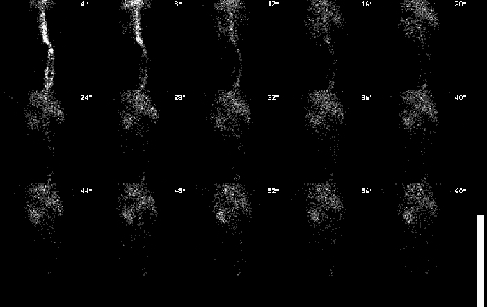
Renal flow images
View main image(rs) in a separate image viewer
View second image(rs).
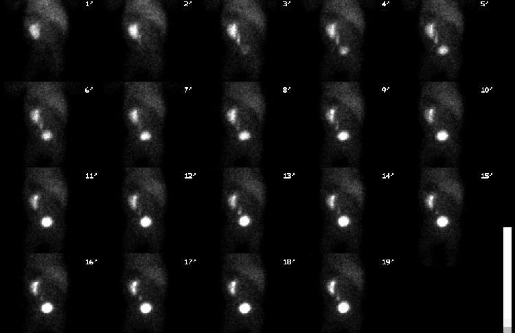
Renal function images
View third image(rs).
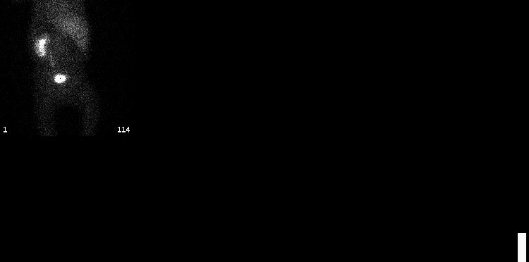
Renal post-void image
Full history/Diagnosis is available below
Diagnosis: Renal Vein Thrombosis
Full history:
6-week-old girl who was admitted to an outside hospital with severe dehydration. The patient was discharged but was readmitted to Barnes-Jewish Hospital with renal failure.
Radiopharmaceutical:
Tc-99m MAG3
Findings:
The posterior abdominal radionuclide venogram demonstrates activity within the suprarenal inferior vena cava. Two vessels of smaller caliber are seen in the infrarenal portion the abdomen and may represent venous collaterals rather than inferior vena cava.
There is perfusion of the left kidney. No perfusion is demonstrated within the right kidney whose position correlates with a photopenic area on the flow images. Sequential renal images show the left kidney to be of normal size. The right kidney is not visualized. There is prompt uptake and excretion of the radiopharmaceutical by the left kidney. There is a small photopenic area in the upper pole of the left kidney which may be related to the presence of the stomach but correlates very well with the area of hypoperfusion demonstrated ultrasonographically. The right kidney is not functional. No abnormalities are seen within the left ureter and the urinary bladder. The right ureter is not visualized.
Discussion:
Renal vein thrombosis is an unfortunate complication of dehydration in a child. A venographic phase of the renal scan is particularly useful in a patient in whom venous thrombosis is suspected. As demonstrated by the early flow images, flow to the infrarenal vena cava is diminished or possibly only present in collaterals suggesting thrombosis or dissection. Dissection is more likely in patients with recent intravascular intervention. The flow images also demonstrate a lack of perfusion to the right kidney.
Absent flow to the right kidney in and of itself may not be a finding with negative implications. The patient may have had a prior nephrectomy or may have an ectopic or congenitally absent kidney. However, in combination with the flow abnormalities identified in the infrarenal IVC, renal venous thrombosis is the prime consideration.
The focal area of decreased perfusion noted on the renal scan also correlates with an area of increased echogenicity on the ultrasound. Differential considerations include an area of prior infarction or previous infection with resultant infarction and scarring.
Followup:
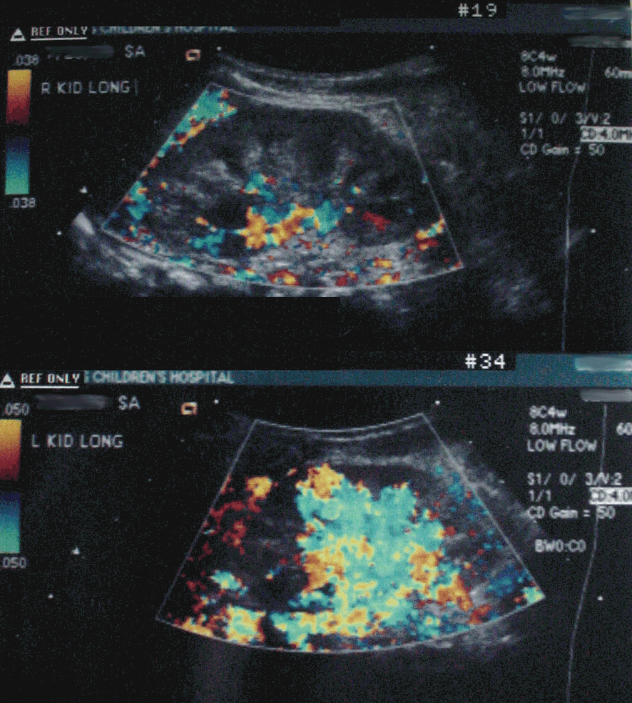
The patient went on to renal ultrasound with color doppler imaging. This revealed thrombus in the right renal vein and infrarenal IVC with resultant decreased perfusion to the right kidney. Also noted was an area of decreased perfusion to the upper pole of the left kidney matching the renal perfusion abnormality.
View followup image(us).
Right (above) and Left (below) Renal Color Doppler Images
Major teaching point(s):
1. The importance of performing and evaluating a venous flow phase of a renal scan
2. The appearance of renal vein thrombosis
3. The appearance of thrombus in the IVC
4. Differential diagnosis of an absent kidney on renal scan
Differential Diagnosis List
1. Renal vein thrombosis
2. Renal artery dissection
3. Fibromuscular dysplasia
ACR Codes and Keywords:
References and General Discussion of Renal Scintigraphy (Anatomic field:Genitourinary System, Category:Organ specific)
Search for similar cases.
Edit this case
Add comments about this case
Return to the Teaching File home page.
Case number: rs024
Copyright by Wash U MO