Case Author(s): Jeffrey Yu, M.D. and Farrokh Dehdashti, M.D. , 1/24/2000 . Rating: #D2, #Q3
Diagnosis: Renal Transplant ATN and Urine Leak
Brief history:
Patient status post cadaveric renal transplant with persistently elevated creatinine.
Images:

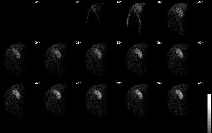
Flow Images
View main image(rs) in a separate image viewer
View second image(rs).
Renal scan sequential images
View third image(mc).
Renal function curve
View fourth image(rs).
Obliques and Post-Void image
Full history/Diagnosis is available below
Diagnosis: Renal Transplant ATN and Urine Leak
Full history:
43 year old man with a history of horseshoe kidney and end stage renal disease. The patient started hemodialysis back in 1975, and had a cadaveric renal transplant in 1977. However, the patient had to re-start dialysis in March, 1998. He had his second cadaveric renal transplant on 9-22-99; however, post-transplant, his creatinine has remained elevated and has increased to 2.8.
Radiopharmaceutical:
Tc-99m MAG3
Findings:
The anterior pelvic radionuclide angiogram demonstrates prompt perfusion of the transplanted kidney in the left iliac fossa. The initial images demonstrate normal transplant size, morphology, and tracer accumulation. The sequential images and renal function curves show prompt uptake and excretion of radiopharmaceutical into the collecting system, but paranchymal retention of the radiopharmaceutical by the transplant kidney.
In the left mid ureter, a focal area of extravasation is identified starting at approximately 6 minutes of imaging. This demonstrates progressive accumulation. The collection tracks along the lateral aspect of the kidney. The patient had a Foley catheter in place. Activity is seen in bladder and the Foley bag.
Discussion:
A persistently elevated creatinine in a post-transplant patient is a worrisome finding that deserves further investigation. Differential diagnosis prior to imaging includes renal vascular compromise, renal infarction, acute rejection, acute tubular necrosis, compression of the kidney or vascular supply by a lymphocele, obstruction, or urine leak.
Renal scintigraphy can be very helpful in narrowing the differential list as it allows one to evaluate renal perfusion, morphology, excretion, as well as the intactness of the renal collecting system, ureters, and bladder.
In this patient, the transplanted kidney was well perfused as documented by the serial images and the renal function curves thereby excluding vascular compromise from the differential list. Similarly, the normal morphology of the kidney makes lymphocele or infarct unlikely. However, the delayed excretion of the tracer into the collecting system suggesting ATN.
While ATN alone could be the cause of the patient's elevated creatinine, close examination of the images reveal a focal collection of tracer along the mid-ureter starting at the 6 minute image. Arrows highlight this area on the follow-up image below. The collection progressively increased in size with time and is suggestive of a urine leak.
So, in this patient it turns out that there were two causes for the persistently elevated creatinine.
Followup:
The patient went to the operating room and initially there was no obvious sign of leakage. However, given the highly suspicious findings on the renal scan, the surgeons decided to inject a dilute solution of dye into the renal pelvis. This revealed a tiny hole at the junction of the renal pelvis and proximal ureter. In retrospect, there was a possibility of ureteral injury at the time of transplant and it is thought that the ureter may have been injured at this site during the transplant operation. This site was closed with a pursestring. Post surgery, the patient's creatinine slowly returned to the normal range.
View followup image(rs).
Sequential renal images annotated with arrows highlighting the urine leakage site.
Major teaching point(s):
1) Renal scans can be very helpful in the evaluation of patients with post-transplant cretinine elevation
2) It is important to assess renal perfusion, uptake, morphology, function, as well as looking closely for sites of urine leakage.
Differential Diagnosis List
1) Renal vascular compromise
2) Renal infarction
3) Acute rejection
4) Acute tubular necrosis
5) Compression of the kidney or vascular supply by a lymphocele
6) Obstruction
7) Urine leak.
ACR Codes and Keywords:
References and General Discussion of Renal Scintigraphy (Anatomic field:Genitourinary System, Category:Effect of Trauma)
Search for similar cases.
Edit this case
Add comments about this case
Return to the Teaching File home page.
Case number: rs023
Copyright by Wash U MO

