Case Author(s): Tate Allen, M.D. and Farrokh Dehdashti, M.D. , 9/28/98 . Rating: #D2, #Q4
Diagnosis: Renal Duplication with Ectopic Ureterocele
Brief history:
Antenatal right hydronephrosis
Images:
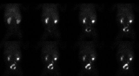
Renal Scintigraphy
View main image(rs) in a separate image viewer
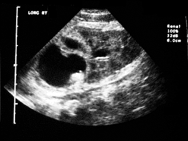
View second image(us).
Right Renal Ultrasound
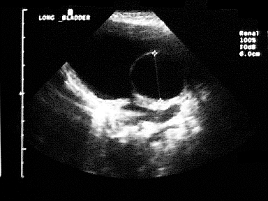
View third image(us).
Bladder Ultrasound
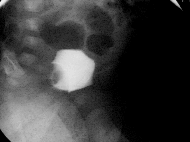
View fourth image(fl).
VCUG
Full history/Diagnosis is available below
Diagnosis: Renal Duplication with Ectopic Ureterocele
Full history:
This is a two-month-old female with a duplicated right kidney.
Radiopharmaceutical:
1.1 mCi Tc-99m MAG3 i.v.
Findings:
The renal scan shows normal perfusion to the kidneys. There is normal
uptake and excretion by the left kidney and the lower pole of the right
kidney. There is delayed uptake and no excretion of the radiopharmaceutical
within the upper pole of the right kidney. This is constistant with
obstruction of the upper pole. An ultrasound of the right
kidney (second image) shows a dilated upper pole collecting system. The
renal scintigram also has a large filling defect within the
urinary bladder. An ultrasound of the bladder (third image) and a VCUG
(fourth image) demonstrate this to represent an ectopic ureterocele.
Discussion:
Complete renal duplication is more common in girls than boys. The lower
pole moiety inserts in the normal trigone location. The upper
pole moiety inserts ectopically which is medial and caudal to the normal
ureteric orifice. The is known as the Weigert-Meyer rule. The upper
pole is often obstructed and is almost always associated with a
ureterocele.
ACR Codes and Keywords:
- General ACR code: 81
- Genitourinary System:
8.14 "CONGENITAL ANOMALY, DEVELOPMENTAL ABNORMALITY exclude: of fetus (.825, .87)"
References and General Discussion of Renal Scintigraphy (Anatomic field:Genitourinary System, Category:Normal, Technique, Congenital Anomaly)
Search for similar cases.
Edit this case
Add comments about this case
Read comments about this case
Return to the Teaching File home page.
Case number: rs019
Copyright by Wash U MO