Case Author(s): John R. Leahy, M.D. and Henry D. Royal, M.D. , 8/9/98 . Rating: #D2, #Q3
Diagnosis: Transplant infarction
Brief history:
49 year old woman with anuria soon after renal transplant.
Images:

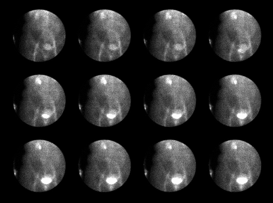
Anterior flow images at 4 sec/frame after 8 sec delay.
View main image(rs) in a separate image viewer
View second image(rs).
Anterior images at 4 min/frame after 3 min delay.
View third image(us).
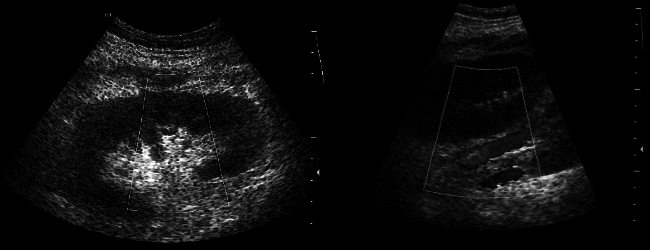
Renal ultrasound with color doppler.
Full history/Diagnosis is available below
Diagnosis: Transplant infarction
Full history:
49 year old woman with end stage renal disease underwent a living related
donor transplant to the right iliac fossa. She developed anuria one week
later.
Radiopharmaceutical:
8.2 mCi Tc-99m MAG3 i.v.
Findings:
The anterior pelvic radionuclide angiogram demonstrates no perfusion of
the transplanted kidney in the right iliac fossa. On delayed images,
there is no appreciable tracer uptake and no excretion from the transplant.
Minimal activity is seen within the patient's native kidneys and bladder.
Renal ultrasound performed the same day shows normal kidney morphology
and normal gray scale appearance. No flow was detected within the renal
artery.
Discussion:
The likely causes of renal transplant dysfunction change depending
on the length of time after surgery. In the immediate post-operative
period, these causes include acute tubular necrosis (ATN), hyperacute
or accelerated rejection, urine leak, hematoma, and infection.
Vascular thrombosis can occur at any time, while arterial stenosis tends
to occur after the first month. Clinically, ATN, rejection, cyclosporin
toxicity and vascular thrombosis all result in decreased renal function.
Treatment of renal dysfunction depends on its cause.
Imaging can help determine the cause of the renal dysfunction.
Renal scintigraphy allows assessment of both perfusion and
function in the transplanted kidney. Activity should be seen in the
kidney 3-6 seconds after activity in the iliac artery.
Peak activity in the
graft should occur in less than 5 minutes, followed by prompt wash-out.
In ATN, there is a slight reduction in perfusion, with significant
parenchymal dysfunction. In rejection, there is decreased perfusion
and relatively preserved function early, with reduction in both
perfusion and function with more chronic rejection. In practice, it is usually not possible
to definitively distinguish ATN from rejection based on the scintigraphic
findings because there is considerable overlap of the findings for these
two causes of renal dysfunction. In arterial thrombosis, there is severe or
total reduction in perfusion and function.
References:
Sandler MP et al: Diagnostic Nuclear Medicine, 3rd ed. Baltimore, Williams
and Wilkins 1996. 1223-29, 1331-39.
Dubovsky EV, Russell CD: Radionuclide evaluation of renal transplants.
Semin in Nucl Med 1988; 18: 181-198
Dodd GD, Tublin ME, Shah A: Vascular complications associated with renal
transplants. AJR 1991; 157:449-459
Major teaching point(s):
Renal scintigraphy and renal ultrasound are complimentary modalities for
use in evaluating the transplanted patient. Ultrasound can confirm
absence of renal arterial flow, and possibly show the location
of the thrombus.
ACR Codes and Keywords:
- General ACR code: 84
- Genitourinary System:
8.455 "Renal transplant, complication of transplant"
References and General Discussion of Renal Scintigraphy (Anatomic field:Genitourinary System, Category:Effect of Trauma)
Search for similar cases.
Edit this case
Add comments about this case
Return to the Teaching File home page.
Case number: rs018
Copyright by Wash U MO