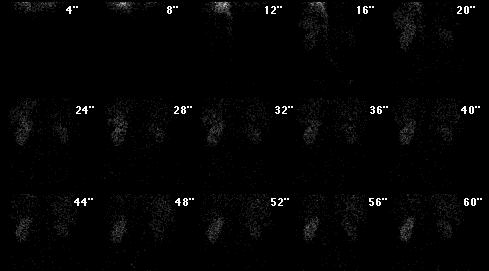
Posterior radionuclide angiogram
View main image(rs) in a separate image viewer
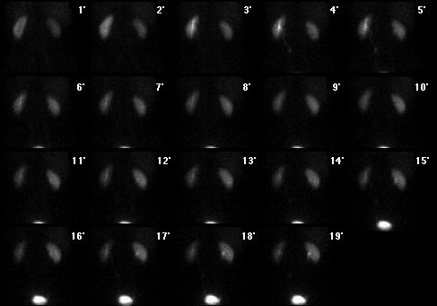
View second image(rs). Posterior sequential images
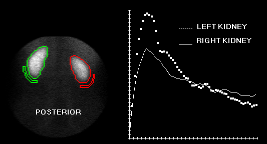
View third image(mm). Split renal function ROI's (on left); Time-activity curves (on right)
Full history/Diagnosis is available below
The estimated contribution of the right kidney to total renal function is 30% and that of the left kidney is 70%.
These scintigraphic findings in a patient with no reported history of renal parenchymal disease are most consistent with renin-mediated hypertension due to right renal artery stenosis.
In patients with renovascular hypertension, compensatory mechanisms via the renin-angiotensin system occur. ACE inhibitors inhibit the conversion of angiotensin I to angiotensin II, which is a potent vasoconstrictor. In patients with renovascular hypertension, this effect results in loss of vasoconstriction of the efferent arterioles which is necessary to maintain effective filtration pressure. Consequently, a decrease in renal function occurs which can be detected scintigraphically.
In patients with renovascular hypertension, ACE inhibitor renal scintigraphy performed with a primarily tubular agent such as Tc-99m MAG3 will classically demonstrate preservation of initial uptake and excretion of tracer. However, there is prolonged retention of parenchymal activity due to the reduced GFR. Initial cortical uptake of Tc99m MAG3 may be decreased in severe stenoses. If a glomerular filtration agent such as Tc99m DTPA is employed, poor initial cortical uptake of tracer is noted predominantly, and prolonged parenchymal retention of activity may also occur. Perfusion to the involved kidney is typically preserved with both renal tubular and glomerular filtration agents, although in severe renal artery stenosis, perfusion to the affected kidney may be diminished.
Patients who are on ACE inhibitors for blood pressure control should stop their medication for 1-3 days, depending on the actual ACE inhibitor, prior to the examination. ACE-inhibitor renal scintigraphy can be performed using a one-day, two-stage protocol, or a one-stage protocol. With a one-stage protocol, renal scintigraphy with ACE inhibition is performed first, and if normal, no baseline study need be performed and renovascular hypertension is excluded. This is usually done in patients with no previous history of renal dysfunction. With respect to the case presented, the ACE-inhibitor renal scintigraphy was so strikingly abnormal that the findings were highly suspicious for renin-mediated hypertension due to right renal artery stenosis. With a one-day, two-stage protocol, a baseline study is first performed followed by a repeat examination with ACE-inhibition.
Following right renal artery angioplasty and stent placement, the patient's blood pressure initially decreased to the normal range, but then intermittently increased. The patient returned for a follow-up ACE-inhibior renal scintigraphy study to evaluate for recurrent renovascular hypertension. The study (not shown) demonstrated significant interval improvement in right renal flow, uptake, and excretion of tracer as well as improved relative renal function since the previous study, indicating successful restoration of blood flow to the right kidney following renal artery angioplasty and stent placement. The split renal function was now more equal, with the right kidney contributing 41% and the left kidney 59% to total renal function. There was no evidence to suggest renin-mediated hypertension.
Reference: Mettler FA and Guiberteau MJ. Essentials of Nuclear Medicine Imaging. 4th ed. 1998. WB Saunders Company. Philadelphia, 349-354.
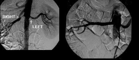
View followup image(an). Renal arteriogram (anterior view) on left; Post angioplasty of and placement of stents in right renal artery on right
References and General Discussion of Renal Scintigraphy (Anatomic field:Genitourinary System, Category:Other generalized systemic disorder)
Return to the Teaching File home page.