Case Author(s): Anton J. Johnson, M.D., Ph.D. and Henry D. Royal, M.D. , 1/29/97 . Rating: #D2, #Q5
Diagnosis: Renal cell carcinoma
Brief history:
49 year old male with back pain.
Images:

Renal Scintigraphy
View main image(rs) in a separate image viewer
Full history/Diagnosis is available below
Diagnosis: Renal cell carcinoma
Full history:
49 year old male with recently diagnosed renal cell carcinoma involving
his right kidney. Renal scintigraphy was requested to evaluate left
renal function. The patient has known metastatic disease to bone.
Radiopharmaceutical:
7.8 mCi Tc-99m MAG3 i.v.
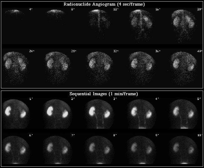
Findings:
The posterior abdominal radionuclide angiogram demonstrates increased blood flow to the upper pole of the right kidney
corresponding to the site of the patient's known tumor. There is
otherwise prompt, symmetrical perfusion to the remainder of the right
and left kidneys.
Two additional sites of increased blood flow and blood pool are seen outside of the
kidneys -- one at about the level of the L2 vertebral body and the
other at about L4 or L5. Both of these regions correspond to sites of
known metastatic disease to bone. Sequential one minute images
show decreased function in the upper pole of the right kidney. The mid and lower
pole of the right and entire left kidney demonstrate normal uptake and
excretion.
Discussion:
Renal cell carcinoma (RCC) is the most common malignant tumor of renal
origin. It has a peak incidence in the sixth decade, occurs twice as
commonly in males, and has a tendency to metastasize to lungs, bone, and
liver. Although it commonly presents with painless hematuria, RCC can
present with symptoms related to metastatic disease (as in this case).
The vast majority of RCC tumors are hypervascular on contrast enhanced
CT and conventional angiography. The findings on renal scintigraphy
frequently include increased flow on the radionuclide angiogram and
decreased activity on the delayed images. The flow findings are
variable, however, and lack of increased flow should not be used as an
argument against RCC.
Fortunately, front line imaging modalities such as CT, US, and MRI are
much better than renal scintigraphy at characterizing renal tumors. The
value of renal scintigraphy in the workup of renal mass lesions comes
from its ability to provide functional information prior to surgical
intervention.
References: Datz FL: Gamuts in Nuclear Medicine, 3rd ed. St. Louis,
Mosby, 1995, p333. Datz FL: Handbook of Nuclear Medicine, 2nd ed. St.
Louis, Mosby, 1993, p168. Dunnick NR, et al: Textbook of Uroradiology,
Baltimore, Williams & Wilkins, p113. Sandler MP, et al: Diagnostic
Nuclear Medicine, 3rd ed. Baltimore, Williams & Wilkins, pp1201-1202.
View followup image(ct).
Single axial image from a CT study shows a large right renal mass as
well as a metastatic focus in the posterior right twelfth rib near its
vertebral articulation. The anterior midline structure with central low
attenuation is normal stomach.
Differential Diagnosis List
The differential diagnosis for a hypervascular lesion in the region of
the kidney includes renal cell carcinoma, abscess, arteriovenous
malformation, gastric activity simulating a mass, renal metastasis,
pheochromocytoma simulating a renal mass, angiomyolipoma, and prominent
spleen in a normal patient.
ACR Codes and Keywords:
References and General Discussion of Renal Scintigraphy (Anatomic field:Skeletal System, Category:Neoplasm, Neoplastic-like condition)
Search for similar cases.
Edit this case
Add comments about this case
Return to the Teaching File home page.
Case number: rs014
Copyright by Wash U MO

