Case Author(s): Scott Winner, M.D. and Tom R. Miller, M.D., Ph.D. , 01/10/97 . Rating: #D2, #Q4
Diagnosis: Infarcted renal transplant
Brief history:
40-year old woman with a renal transplant in the right iliac fossa.
Images:

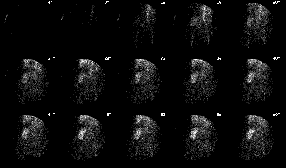
Anterior pelvic radionuclide angiogram four weeks after transplantation
View main image(rs) in a separate image viewer
View second image(rs).
Sequential dynamic images four weeks post-transplant
View third image(rs).
Anterior pelvic radionuclide images two weeks after the first study
View fourth image(rs).
Sequential dynamic images from the second study
Full history/Diagnosis is available below
Diagnosis: Infarcted renal transplant
Full history:
40-year old diabetic female
with end-stage renal disease. She received a
renal transplant and was doint well at the time of the first study. The
patient presented in renal failure on at the time of the second study.
Radiopharmaceutical:
7.6 mCi Tc-99m
MAG3 i.v.
Findings:
The first renal transplant
scintigraphy demonstrates
prompt perfusion to the transplanted kidney
with only mildly delayed uptake and excretion
of tracer. The second study two weeks later (when the patient1s creatinine was
7.4) demonstrates no appreciable perfusion,
uptake, or excretion of tracer by the
transplant.
Discussion:
The lack of appreciable perfusion to the transplanted kidney on the second study was strong evidence that the transplant was not perfused and, hence, infarcted. Because of the less likely possibility that the findings were due to severe renal failure,
a sonogram was performed, showing no blood flow to the kidney.
ACR Codes and Keywords:
- General ACR code: 84
- Genitourinary System:
8.481 "Vascular occlusion exclude: in transplanted kidney (.4557)"
References and General Discussion of Renal Scintigraphy (Anatomic field:Genitourinary System, Category:Effect of Trauma)
Search for similar cases.
Edit this case
Add comments about this case
Read comments about this case
Return to the Teaching File home page.
Case number: rs013
Copyright by Wash U MO

