Case Author(s): Brigid Gordon, MD and Henry Royal, MD , 08/04/96 . Rating: #D2, #Q4
Diagnosis: Pyelonephritis
Brief history:
2 year old female with fevers, pyuria, and leukocytosis.
Images:
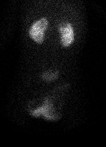
Posterior views of both kidneys using LEAP collimator
View main image(rs) in a separate image viewer
View second image(rs).
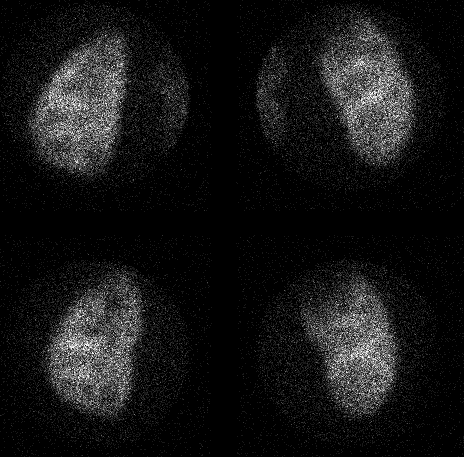
Pinhole images of the kidneys
(Upper row LPO and RPO, lower row posterior views)
Full history/Diagnosis is available below
Diagnosis: Pyelonephritis
Full history:
2 year old female with fever, leukocystosis, and pyuria with negative
blood and urine cultures. No prior documented urinary tract infections.
Because of persistent fevers despite antibiotics, a renal ultrasound was
obtained prior to the renal nuclear scan. The ultrasound suggested a focus
of increased echogenicity in the upper pole of the right kidney, but
without cortical thinning to suggest prior scarring (sonogram
not shown).
Radiopharmaceutical:
TC99m-DMSA
Findings:
Immediate blood flow and static images (not included) demonstrated
decreased flow and tracer uptake to the upper pole of the right kidney.
The posterior images (provided above) were obtained 3 hours after the
injection of the tracer. Both kidneys are normal in size and location.
However, there is a cortical defect in the upper pole of the right
kidney that corresponded to the abnormality on the immediate and static
views.
Discussion:
Pyelonephritis, scar, and solid renal masses can produce decreased uptake
of this radiopharmaceutical. In the case of infection or pyelonephritis,
it is postulated that there is an inactivation of the transport mechanism
for the radiopharmaceutical across the cell membrane into the tubular
cells. This results in decreased localization of the tracer (i.e.
cortical defect) at the site of infection. TC99m-DMSA imaging provides
visualization of the renal parenchyma without interference from retention
of tracer in the collecting system.
Major teaching point(s):
Cortical defects caused by infection.
Differential Diagnosis List
Differential includes both infection, scar and renal masses.
ACR Codes and Keywords:
References and General Discussion of Renal Scintigraphy (Anatomic field:Genitourinary System, Category:Inflammation,Infection)
Search for similar cases.
Edit this case
Add comments about this case
Return to the Teaching File home page.
Case number: rs010
Copyright by Wash U MO