Case Author(s): Jerold Wallis, M.D. , 7/5/96 . Rating: #D1, #Q3
Diagnosis: Normal renal parenchymal imaging study
Brief history:
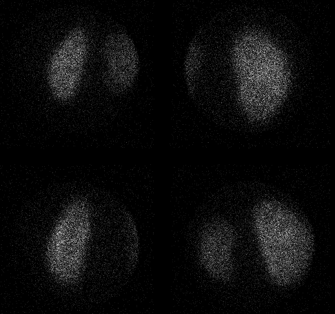
Patient with vesicoureteral reflux, evaluate for renal scarring.
Images:

Above: left and right posterior pinhole images
Below: posterior oblique pinhole images
View main image(rs) in a separate image viewer
View second image(rs).
For comparison,
standard posterior images with a high
resolution low energy collimator.
Full history/Diagnosis is available below
Diagnosis: Normal renal parenchymal imaging study
Full history:
2 year old child being evaluated for renal parenchymal scarring.
Radiopharmaceutical:
0.9 mCi Tc-99m DMSA
Findings:
Normal study. The left kidney is slightly smaller than the right.
Discussion:
Renal imaging with DMSA can demonstrate evidence of
parenchymal scarring due to prior infection, as well
as evidence of focal reaction to infection in the
setting of pylonephritis.
If the object is to detect renal scarring,
the patient must be free of infection for a few
months prior to the exam.
When used for the detection of pylonephritis, the
sensitivity and specificity have been reported to
be better than 95%.
ACR Codes and Keywords:
References and General Discussion of Renal Scintigraphy (Anatomic field:Genitourinary System, Category:Normal, Technique, Congenital Anomaly)
Search for similar cases.
Edit this case
Add comments about this case
Read comments about this case
Return to the Teaching File home page.
Case number: rs009
Copyright by Wash U MO

