Case Author(s): Charles Pringle, M.D., Barry A. Siegel, M.D. , 01/10/96 . Rating: #D2, #Q3
Diagnosis: Vesicoureteral Reflux
Brief history:
Past history of colon cancer
Images:

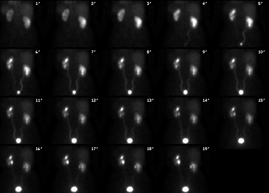
Posterior images, 1 minute through 19
minutes after injection of Tc-99m MAG3
View main image(rs) in a separate image viewer
View second image(rs).
Sequential images after administration of furosemide (Lasix).
View third image(mm).
Split renal function calculation and time-activity curve
View fourth image(mm).
Lasix washout curves
Full history/Diagnosis is available below
Diagnosis: Vesicoureteral Reflux
Full history:
Previous history of colon cancer
approximately 12 years ago. Treatment included
radiation therapy. The patient subsequently
developed radiation cystitis and a small-capacity
bladder. She also subsequently developed bilateral
ureteral obstruction, which has been treated with
bilateral ureteral stents, which are now in place.
Radiopharmaceutical:
7.3 mCi Tc-99m MAG3
Findings:
There is prompt mildly asymmetric
perfusion to both kidneys, with left kidney perfusion
slightly less than the right. There is also prompt
uptake and excretion by both kidneys, although the
left renal excretion is slightly less than the right.
Split renal function is 62% right kidney and 38% left
kidney.
Despite the intravenous administration of furosemide, there
appears to be an increase in radiopharmaceutical
activity within the collecting systems of both kidneys
over time. This is consistent with bilateral reflux into
the ureteral stents and into the collecting systems of
both kidneys. The patient's small capacity bladder
also most likely contributes to this process.
Followup:
Subsequent cystoscopy revealed a
markedly decreased bladder capacity (75 mL total).
Major teaching point(s):
It is importance to correlate
the post-diuretic washout curve with the appearance of the images. In
this case, the images confirmed the vesicoureteral
reflux. This is not unusual in patients with ureteral
stents.
ACR Codes and Keywords:
References and General Discussion of Renal Scintigraphy (Anatomic field:Genitourinary System, Category:Organ specific)
Search for similar cases.
Edit this case
Add comments about this case
Return to the Teaching File home page.
Case number: rs008
Copyright by Wash U MO

