Case Author(s): Hamid Latifi,MD , 4/14/95 . Rating: #D2, #Q3
Diagnosis: Prune belly syndrome
Brief history:
33 year old man with slowly increasing
creatinine.
Images:
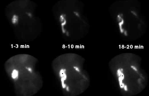
Posterior images of abdomen through 20 minutes. Both rows of images are the same but the bottom row is at higher intensity to show the soft tissues.
View main image(rs) in a separate image viewer
View second image(rs).
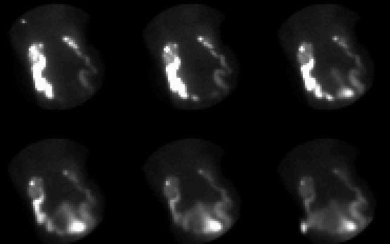
Post-furosemide posterior images of
the abdomen, 5 minutes per frame for
30 minutes.
Full history/Diagnosis is available below
Diagnosis: Prune belly syndrome
Full history:
33-year old man with known diagnosis of prune belly syndrome
and history of multiple prior urinary tract infections
who now presents with a slowly increasing serum creatinine.
The patient has had a prior vesicostomy. Renal scintigraphy
was requested to evaluate relative renal function and exclude
renal obstruction.
Findings:
Renal scintigraphy shows a small right kidney with normal
left renal function. The collecting systems are dilated
bilaterally, worse on the left. The ureters are laterally
deviated, which is not unusual in patients with prune belly
syndrome. There is no evidence of urodynamically significant
obstruction, as documented by prompt clearance of activity
from both renal collecting systems after administration of
intravenous furosemide.
Discussion:
This case simply demonstrates a renal scintigraphic
appearance of an adult patient with prune belly syndrome.
ACR Codes and Keywords:
References and General Discussion of Renal Scintigraphy (Anatomic field:Genitourinary System, Category:Normal, Technique, Congenital Anomaly)
Search for similar cases.
Edit this case
Add comments about this case
Read comments about this case
Return to the Teaching File home page.
Case number: rs006
Copyright by Wash U MO