Case Author(s): Delphine L. Chen, M.D., and Farrokh Dehdashti, M.D. , 6/29/06 . Rating: #D3, #Q4
Diagnosis: Hodgkin's lymphoma response to therapy
Brief history:
52 yo. man diagnosed with Hodgkin's lymphoma who has completed 2 cycles of chemotherapy.
Images:

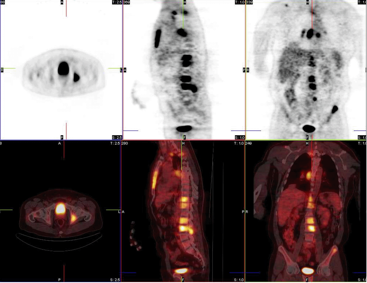
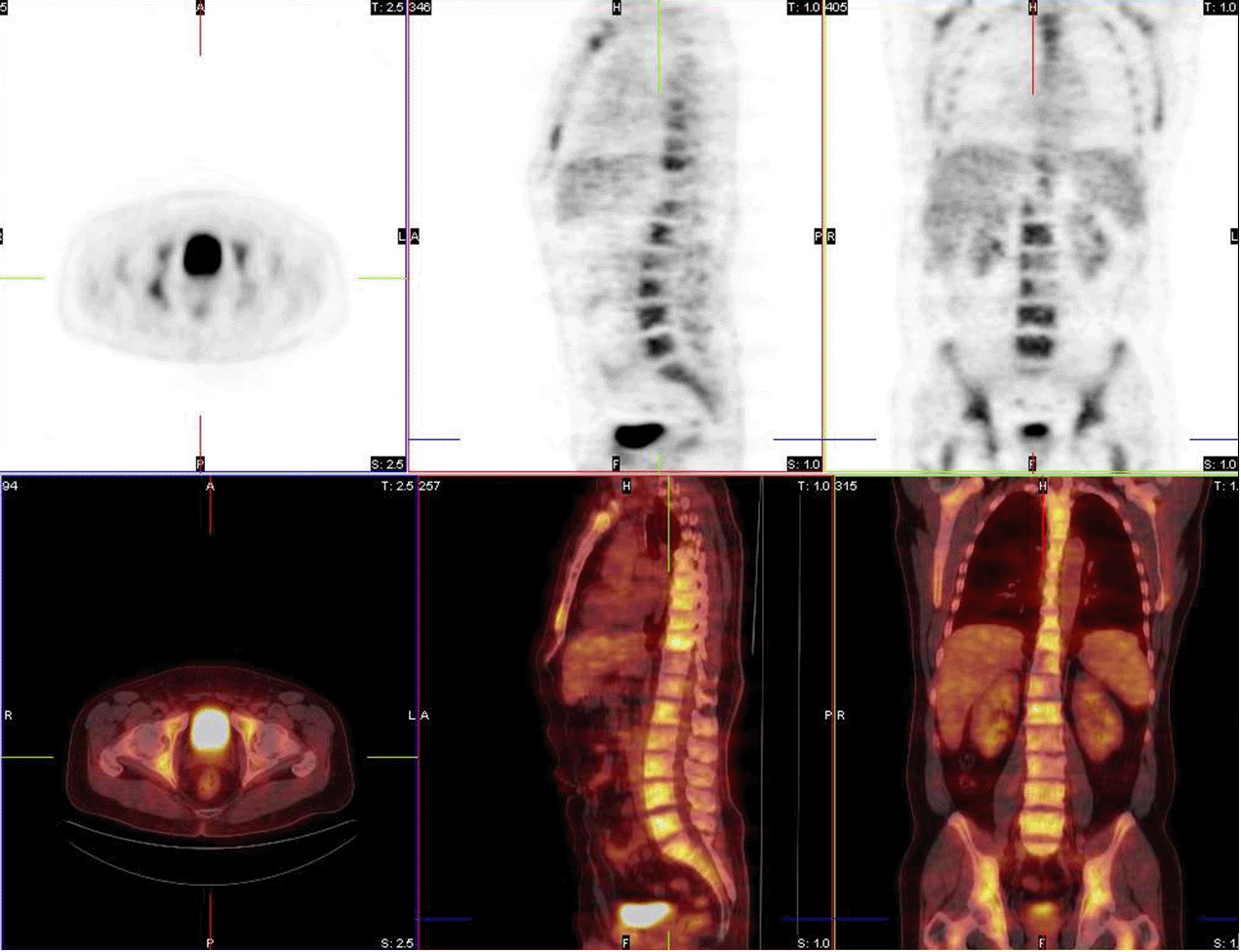
Anterior projections from MIP images at initial diagnosis and after 2 cycles of chemotherapy.
View main image(pt) in a separate image viewer
View second image(pt).
Fusion images before treatment.
View third image(pt).
Fusion images after 2 cycles of chemotherapy.
View fourth image(pt).
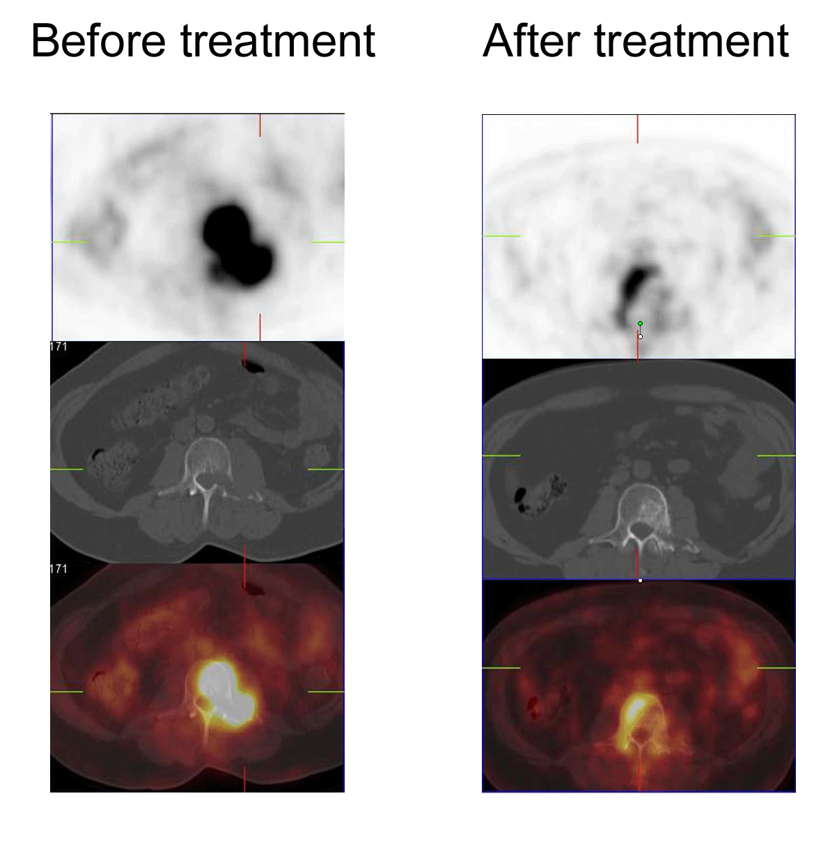
Vertebral body lesion before and after 2 cycles of chemotherapy.
Full history/Diagnosis is available below
Diagnosis: Hodgkin's lymphoma response to therapy
Full history:
52 yo. man with Hodgkin's lymphoma who had received 2 cycles of ABVD chemotherapy. PET/CT imaging is requested for treatment monitoring.
Radiopharmaceutical:
14.8 mCi F-18 fluorodeoxyglucose i.v.
Findings:
PET-CT at initial diagnosis demonstrated multiple areas of bulky lymphadenopathy in the neck and chest, adenopathy in the abdomen, and multiple skeletal lesions, including the spine and pelvis.
PET-CT after 2 cycles demonstrated significantly reduced FDG uptake in all soft tissue and bony lesions. Additionally, because of recent granulocyte colony stimulating factor administration in association with chemotherapy, there is intense FDG uptake in the normal bone marrow, accentuating areas of lymphomatous involvement in the skeleton, which now have decreased uptake. Most bony lesions on CT demonstrated no change in degree of sclerosis, but a few demonstrated increased sclerosis (see 4th image above).
Discussion:
PET imaging with FDG has been demonstrated to be much more sensitive and specific than CT in detecting residual disease in lymphoma after treatment, since CT cannot be used to distinguish between tumor and fibrosis in residual masses (note in this case that few of the bony lesions had significantly changed in appearance on CT, but all demonstrated decreased FDG uptake). A growing body of evidence also suggests that using FDG-PET imaging early after initiation of chemotherapy may predict which patients are more likely to relapse; several studies have demonstrated that a negative PET study in patients with Hodgkin's disease has high negative predictive value and that patients with negative PET studies after completing treatment have longer progression-free survival than those with residual disease on PET (Kumar 2004).
Additionally, several small studies have demonstrated that early imaging with FDG-PET, after 2-3 cycles of chemotherapy, may be useful in predicting which patients are more likely to relapse within a short period of time (Kumar 2004). Those patients with persistent FDG uptake after 2 cycles tended to recur more quickly after completing chemotherapy, whereas most patients with no FDG uptake tended to have prolonged progression-free periods (Kumar 2004).
Reference: Kumar R, Maillard I, Schuster SJ, Alavi A. Utility of fluorodeoxyglucose-PET imaging in the management of patients with Hodgkin's and non-Hodgkin's lymphomas. Radiologic Clinics of North America. 2004;42(6):1083-100.
Followup:
This case is a dramatic example of the marked response to chemotherapy that can be seen on FDG-PET scans obtained early in the course of treatment in patients with lymphoma. There was little to no residual FDG uptake seen in the areas of lymphomatous involvement on the treatment monitoring PET scan. The patient completed his chemotherapy course and had no evidence of recurrent disease by PET-CT at one year after diagnosis.
ACR Codes and Keywords:
References and General Discussion of PET Tumor Imaging Studies (Anatomic field:Skeletal System, Category:Neoplasm, Neoplastic-like condition)
Search for similar cases.
Edit this case
Add comments about this case
Return to the Teaching File home page.
Case number: pt155
Copyright by Wash U MO