Case Author(s): Jabi Shriki, M.D., Rick Held, M.D., and Farrokh Dehdashti, M.D. , 06/29/06 . Rating: #D2, #Q.
Diagnosis: Esophageal carcinoma, upstaged by PET imaging.
Brief history:
52-year-old man with recently diagnosed esophageal cancer at the gastroesophageal junction. FDG-PET/CT study is requested for initial staging.
Images:

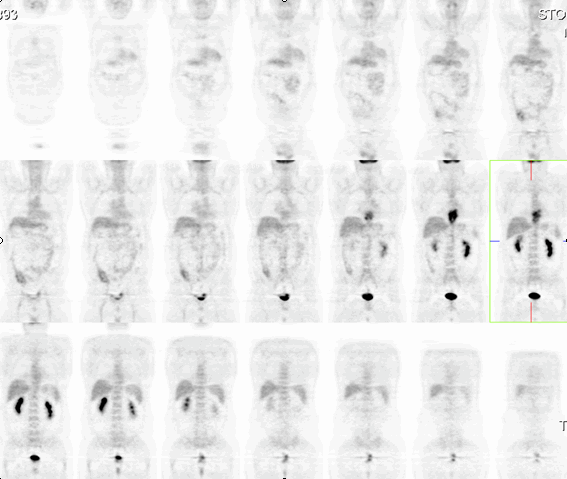
Coronal tomographic images after FDG administration are shown.
View main image(pt) in a separate image viewer
View second image(pt).
Fused, PET/CT images.
Full history/Diagnosis is available below
Diagnosis: Esophageal carcinoma, upstaged by PET imaging.
Full history:
This is a 52-year-old male with newly diagnosed esophageal carcinoma. He presented approximately two weeks previously with chest pain with radiation to the back pain. His pain was associated with dysphagia and dyspepsia. A cardiac work-up was performed and was essentially negative. Subsequently a barium swallow (not shown) was performed and he was found to have a mass at the gastroesophageal junction. Esophagogastroduodenoscopy and biopsy showed an adenocarcinoma of the distal esophagus.
Radiopharmaceutical:
2-deoxy-(2,2)18F-fluoro-D-glucose (FDG)
Findings:
The distal esophageal mass, consistent with the known carcinoma in this region is visualized. Also, a 1.4 by 1.6 cm, periesophageal lymph node is seen, with increased uptake of FDG suggesting nodal disease.
Discussion:
Esophageal carcinomas are staged with a traditional, TNM staging system. However, unlike many other gastrointestinal tumors, esophageal nodal staging is divided into N0 (no nodal involvement) and N1 (locoregional lymph node involvement) only. Once there is spread outside of locoregional nodes, an M1 status is designated. The presence of N1 nodal disease upstages esophageal carcinoma to at least a stage IIB tumour, and decreases the expected 5-year survival from 40% to 20%. Although the mainstays of staging of esophageal carcinoma continue to be conventional computed tomography and endoscopic ultrasound, and important role of FDG-PET/CT is evolving.
References:
http://www.emedicine.com/radio/topic271.htm
Jadvar H, Henderson RW, Conti PS. Mol Imaging Biol. 2006 May-Jun;8(3):193-200.
Followup:
Because of concomitant coronary disease, this patient was felt to be a poor surgical candidate, and was therefore, placed on a study protocol of chemoradiation. Unfortunately, his disease subsequently progressed on a repeat PET/CT scan (not shown) while on chemoradiation.
Major teaching point(s):
The importance of FDG-PET/CT in characterizing lymph nodes in esophageal cancer.
Differential Diagnosis List
Metastatic vs. inflammatory lymph nodes.
ACR Codes and Keywords:
References and General Discussion of PET Tumor Imaging Studies (Anatomic field:Gasterointestinal System, Category:Neoplasm, Neoplastic-like condition)
Search for similar cases.
Edit this case
Add comments about this case
Return to the Teaching File home page.
Case number: pt154
Copyright by Wash U MO

