Case Author(s): Xia Wang, M.D. and Barry A. Siegel, M.D. , . Rating: #D2, #Q3
Diagnosis: Left Buttock Large B-cell Lymphoma.
Brief history:
41-year-old man with a left buttock mass
Images:

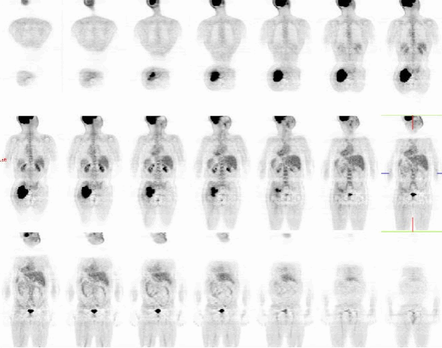
Prone coronal PET/CT images
View main image(pt) in a separate image viewer
View second image(pt).
Prone axial PET/CT images
View third image(pt).
3 views PET/CT Prone Images.
Full history/Diagnosis is available below
Diagnosis: Left Buttock Large B-cell Lymphoma.
Full history:
41-year-old man with a left buttock mass. Biopsy showed large B-cell lymphoma. This is an initial staging examination.
Radiopharmaceutical:
15.0 mCi F-18 Fluorodeoxyglucose i.v.
Findings:
There is markedly increased FDG uptake within a large mass in the left buttock musculature, which extends through the sciatic notch and into the left mid pelvis. The maximum standardized uptake value of the mass is 23.5. The mass demonstrates heterogeneity in its FDG uptake. There is no other focus of abnormal FDG uptake noted on this examination.
The study was done in the prone position, because the mass prevented the patient from lying comfortably in the supine position.
Discussion:
FDG PET is increasingly used to monitor tumor response in patients undergoing chemotherapy and chemoradiotherapy. Numerous studies have shown that FDG PET is an accurate test for differentiating residual viable tumor tissue from therapy-induced fibrosis. Furthermore, quantitative assessment of therapy-induced changes in tumor FDG uptake may allow the prediction of tumor response and patient outcome very early in the course of therapy. Early identification of chemotherapy-refractory lymphoma patients provides a basis for alternative treatment strategies. Thus, FDG PET has an enormous potential to reduce the side effects and costs of ineffective therapy. However, the optimal timing for imaging during and after treatment has not been established yet.
Reference:
Wolfgang A. Weber, Use of PET for Monitoring Cancer Therapy and for Predicting Outcome, J. Nucl. Med. 2005 46: 983-995.
Lale Kostakoglu, Morton Coleman, John P. Leonard, et al., PET Predicts Prognosis After 1 Cycle of Chemotherapy in Aggressive Lymphoma and Hodgkin’s Disease , J. Nucl. Med. 2002 43: 1018-1027.
Followup:
4 months later, after completion of 6 cycles of CHOP chemotherapy. There has been marked decrease in size and activity of the soft tissue mass in the left gluteal region. There is now atrophy of left gluteus muscles on the corresponding CT images. The maximum standardized uptake value is now 2.2. The findings indicate a successful response to treatment (complete "metabolic" response). There is minimally increased residual activity between the pyriformis muscle and the gluteal muscles.
View followup image(pt).
Post Treatment Axial PET/CT Image.
Major teaching point(s):
FDG PET is useful in monitoring lymphoma response to chemotherapy and chemoradiotherapy.
Differential Diagnosis List
1. Primary: Lymphoma, malignant fibrous histiocytoma, sarcoma (non- specific, such as rhabdomyosarcoma)
2. Metastasis
3.Nonneoplastic processes: infection, myositis, or hematoma.
ACR Codes and Keywords:
References and General Discussion of PET Tumor Imaging Studies (Anatomic field:Skeletal System, Category:Neoplasm, Neoplastic-like condition)
Search for similar cases.
Edit this case
Add comments about this case
Return to the Teaching File home page.
Case number: pt152
Copyright by Wash U MO

