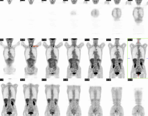
Coronal FDG-PET Imaging.
View MIP cine in AVI format.
View main image(pt) in a separate image viewer
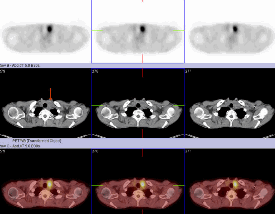
View second image(pt). Axial PET/CT Imaging.
Full history/Diagnosis is available below
There is a focal area of increased FDG uptake in the left lobe of the thyroid, which correlates with a predominantly solid and heterogeneous low attenuating nodule on corresponding CT images. In addition, a faint area of increased uptake is noted just adjacent to the surgical clip in the left axilla. No other areas of abnormally increased FDG uptake are identified.
The incidence of new thyroid lesions found on routine FDG-PET has been previously reported to be 2.2 and 2.3% in two large studies of over a total 4100 oncology patients. Of thyroid lesions that underwent biopsy in these two studies, 50% and 47% were found to be malignant, respectively (1, 2). Thus, 18F-FDG PET is useful for detecting malignant lesions of the thyroid gland.
Focal FDG uptake has also been reported in hyperplastic nodules of the thyroid. 18F-FDG uptake was also significantly higher in Graves’ disease when compared with normal thyroids. Chronic follicular Hashimoto’s thyroiditis usually has diffusely increased FDG uptake but focal uptake can also be seen. Chronic thyroiditis can have a high SUV in the malignant range.
18F-FDG PET is not generally used to differentiate benign from malignant thyroid tumors because there is considerable overlap the SUVs of benign and malignant thyroid tumors. Given the high prevalence of malignancy, patients with focal thyroid uptake should have an FNA to rule out cancer.
Reference:
1. T.Y. Kim, W.B. Kim and J.S. Ryu et al., 18F-fluorodepxyglucose uptake in thyroid from positron emission tomogram(PET) for evaluation in cancer patients: high prevalence of malignancy in thyroid PET incidentaloma, Laryngoscope 115(2005), pp. 1074-1078.
2. M.S. Cohen, N. Arslan and F. Dehdashti et al., Risk of malignancy in thyroid incidentalomas identified by fluorodeoxyglusoce-positron emission tomography, Surgery 130 (2001), pp. 941-946.
3. A.G. Gianoukakis, M. Karam and A. Cheema, et al., Autonomous thyroid nodules visualized by positron emission tomography with 18F-fluorodeoxyglucose: a case report and review of the literature, Thyroid 13 (2003), pp. 395-399.
4. A.R. Boerner, E. Voth and P. Theissen, et al., Glucose metabolism of the thyroid in Graves’ disease measured by F-18-fluoro-deoxyglucose positron emission tomography, Thyroid 8(1998), pp. 765-772.
References and General Discussion of PET Tumor Imaging Studies (Anatomic field:Face, Mastoids, and Neck, Category:Neoplasm, Neoplastic-like condition)
Return to the Teaching File home page.