Case Author(s): Richard Held, M.D. and Barry A. Siegel, M.D. , 06/21/06 . Rating: #D4, #Q5
Diagnosis: Sigmoid colon adenocarcinoma with tumor invasion of the inferior mesenteric vein
Brief history:
76-year-old woman complaining of abdominal pain and diarrhea, as well as increasing confusion. She had imaging performed at another institution, which revealed a cerebellar mass. Surgical resection of the mass was performed, revealing adenocarcinoma of uncertain primary. Further imaging demonstrates a heterogeneous adnexal mass. PET/CT was requested for further staging and diagnosis.
Images:

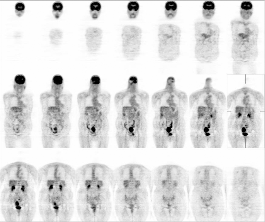
Coronal FDG-PET images
View main image(pt) in a separate image viewer
View second image(pt).
Coronal PET/CT image
View third image(pt).
Axial PET/CT images - PLEASE ALLOW MOUSE TO HOVER OVER IMAGE THEN MOVE TO THE LOWER RIGHT PART OF THE COMPOSITE TO REVEAL THE EXPAND ICON WHICH MAY BE CLICKED FOR BEST ANATOMIC DETAIL.
View fourth image(gs).
Barium enema
Full history/Diagnosis is available below
Diagnosis: Sigmoid colon adenocarcinoma with tumor invasion of the inferior mesenteric vein
Full history:
76-year-old woman who had initially presented to another institution complaining of confusion and abdominal pain. Her abdomincal pain was evaluated with ultrasonography, revealing a complicated right adnexal mass. Then she had a CT scan of her head/chest/abdomen/pelvis for further assessment of suspected malignancy. Her brain CT showed an enchacing cerebellar mass, and the remainder demonstrated a mass in the pelvis, possibly arising from the bowel. She was transferred to this institution for definitive treatment.
She had her cerebellar mass resected. It demonstrated metastatic adenocarcinoma of uncertain origin, but a gastrointestinal primary was favored. She then had a barium enema. It revealed a large, but typical-appearing sigmoid colon carcinoma. CT was then performed and showed a large heterogeneous sigmoid colon mass, adjacent adenopathy, and thrombosis in the inferior mesenteric vein.
PET/CT was requested for staging.
Radiopharmaceutical:
F-18 FDG
Findings:
There is intense FDG uptake within a large mass in the sigmoid colon. The maximum standardized uptake value (SUV max) is 26.4, indicative of malignancy. The size of this lesion is 8.1 x 6.1 cm. There are numerous adjacent mesenteric lymph nodes, which are enlarged and demonstrate intense FDG uptake. In addition, there is linear FDG uptake seen ascending into the inferior mesenteric vein (IMV) and also into a more distal branch point, compatible with invasion of the superior mesenteric vein with tumor thrombus.
Followup:
The patient went to surgery, which confirmed the tumor invasion of the inferior mesenteric vein as detected by PET/CT. Her primary tumor was resected. She is being treated with chemotherapy.
See also the Pre-PET/CT contrast-enhanced CT showing the thrombosed IMV.
View followup image(ct).
Contrast enhanced CT showing the thrombosed inferior mesenteric vein
Major teaching point(s):
Colorectal adenocarcinoma is well known to invade venous structures. Studies that have investigated report a wide range the frequency of venous invasion.
Being aware of this potential mechanism of spread of tumor is important to avoid misdiagnosis. Use of all the clinical information and information and images from previous examinations is also essential in accurate interpretation of any PET/CT study. In this particular case, it was known that the patient had IMV thrombosis on the CT done prior to this examination. It was also known that the patient has a large sigmoid colon tumor with enlarged lymph nodes. What was not known was that the IMV had been invaded by tumor. Clearly, the FDG-PET/CT images show abnormal uptake in the IMV. It is also contiguous with the primary tumor. A search of relevant journal articles reveals several articles citing tumors with different histology, glycolytic rate, infected catheters, and bland thrombus as potentially causing increased venous uptake as well. In this case however, the direct extension from the tumor and degree of FDG uptake allows us to confidently make the diagnosis.
J Clin Pathol. 2002 Jan;55(1):17-21
Clin Nucl Med. 2005 May;30(5):342-3
Diseases of the Colon & Rectum, Volume 34, Issue 9, Sep 1991, Pages 798 - 804
Differential Diagnosis List
Based upon contrast-enhanced CT images (follow-up image 5) the IMV is thrombosed. Differentiation between bland and tumor "thrombus" of the IMV is not possible using standard CT techniques and contrast timing. If possible at all to make the diagnosis on CT, it would require visualization of enhancement within the tumor thrombus to suggest this possibility.
Using PET alone, it would be difficult to correctly identify the IMV, which could easily be mistaken for a ureter or loop of bowel.
PET/CT makes interpretation much easier. The ureter is seen separately from the activity in the IMV, which is not continuous with loops of bowel. The distal branching pattern also identifies it as a vascular structure.
ACR Codes and Keywords:
References and General Discussion of PET Tumor Imaging Studies (Anatomic field:Gasterointestinal System, Category:Neoplasm, Neoplastic-like condition)
Search for similar cases.
Edit this case
Add comments about this case
Return to the Teaching File home page.
Case number: pt148
Copyright by Wash U MO

