Case Author(s): Richard Held, MD and Tom R. Miller, MD, PhD , 6/15/06 . Rating: #D2, #Q3
Diagnosis: Lymphoma (Lymphoblastic Precursor T cell) with pleural involvement and malignant effusion
Brief history:
18-year-old man with a large anterior mediastinal mass. PET/CT study is requested for staging.
Images:

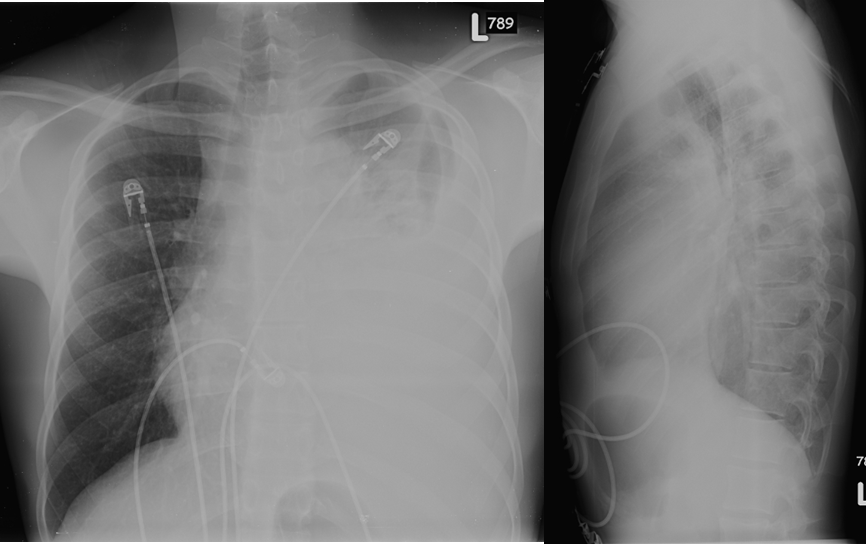
Chest Radiograph
View main image(xr) in a separate image viewer
View second image(pt).
F18-FDG PET - Coronal Images
View third image(pt).
PET/CT axial images with fused and unfused images
View fourth image(ct).
Correlative Chest CT with IV contrast
Full history/Diagnosis is available below
Diagnosis: Lymphoma (Lymphoblastic Precursor T cell) with pleural involvement and malignant effusion
Full history:
This 18-year-old man was admitted for evaluation of a new anterior mediastinal mass. He had a two and one-half month history of mild pleuritic chest pain and a one month history of marked increase in pleuritic chest pain and shortness of breath. A chest radiograph (main image) showed a large anterior mediastinal mass and left pleural effusion. On review of systems, he denies fevers or night sweats. He had a 20-pound weight loss over the last two months.
Radiopharmaceutical:
F-18 FDG
Findings:
The entire pleura of the left lung has intense FDG uptake due to tumor.
There is a large, intensely FDG-avid mass centered in the anterior mediastinum, occupying it nearly completely. This, in conjunction with a large left pleural effusion, causes shift of the mediastinum to the right.
There are multiple associated right retrocrural and left para-aortic lymph nodes. All of these findings demonstrate intense FDG uptake and most likely represent lymphoma with an associated malignant left pleural effusion and metastatic lymphadenopathy.
There are multiple right lung pulmonary opacities with minimal to no FDG uptake; a benign process, such as atelectasis related to rightward shift of the mediastinum, is favored.
Discussion:
This is a somewhat unusual presentation of lymphoma with involvement of the pleura on only one side of the chest and an associated pleural effusion.
Followup:
The patient was treated with chemotherapy for biopsy-proven precursor t-cell lymphoblastic lymphoma.
Major teaching point(s):
There is a limited differential diagnosis for diffuse pleural hypermetabolism: Lymphoma, mesothelioma, empyema/pleurisy and metastatic breast cancer. The presence of an anterior mediastinal mass and retroperitoneal adenopathy strongly favors lymphoma.
Differential Diagnosis List
Lymphoma, mesothelioma, empyema/pleurisy, metastatic breast cancer
ACR Codes and Keywords:
References and General Discussion of PET Tumor Imaging Studies (Anatomic field:Vascular and Lymphatic Systems, Category:Neoplasm, Neoplastic-like condition)
Search for similar cases.
Edit this case
Add comments about this case
Return to the Teaching File home page.
Case number: pt146
Copyright by Wash U MO

