
Coronal PET image. View MIP cine in AVI format.
View main image(pt) in a separate image viewer
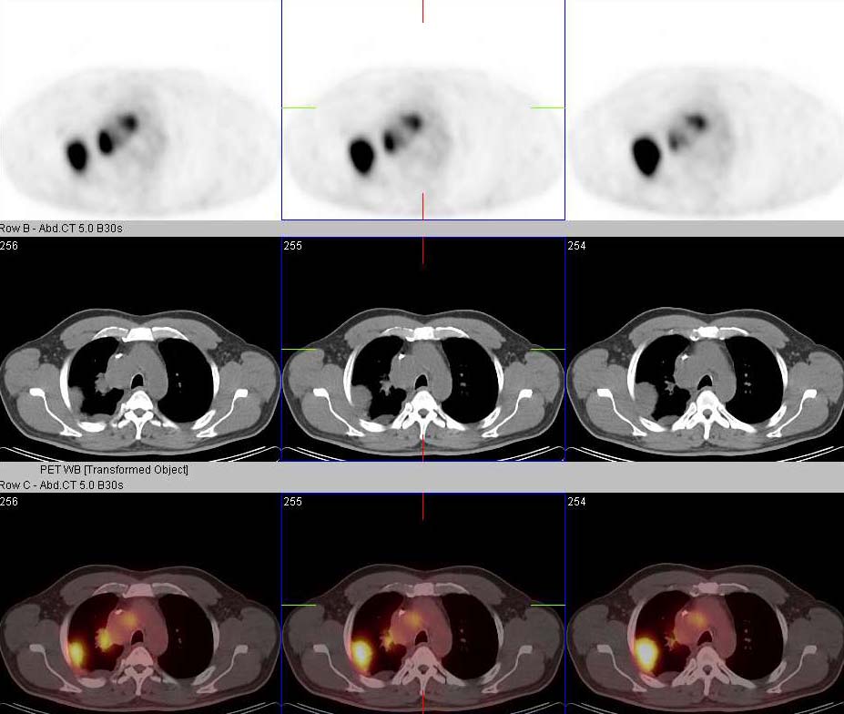
View second image(pt). Axial PET/CT
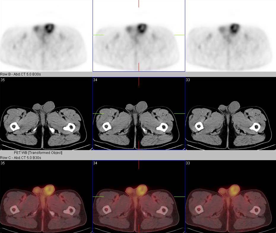
View third image(pt). Axial PET/CT
View fourth image(fl). Chest X-ray
Full history/Diagnosis is available below
14.6 mCi F-18 Fluorodeoxyglucose i.v.
CXR: There are multiple pleural based masses in both lungs and enlarged lymph nodes in the mediastinum.
Tumor markers alpha-fetoprotein (AFP), beta-HCG, and lactate dehydrogenase (LDH) are vital in the evaluation and management of patients with GCTs. They are used for determining diagnosis, staging, prognosis, and response to therapy(1).
GCTs are seen predominantly in whites and rarely in African Americans. GCT is the most common solid tumor in men aged 15-35 years. Risk of developing GCT in the cryptorchid testis is higher than in the normally descended testis(1).
Pattern of metastases is as follows: The right testicular tumors usually metastasize to nodes between the aorta and the inferior vena cava (interaortocaval nodes); left testicular tumors usually metastasize to the pre-aortic and para-aortic nodes (1). Left supraclavicular adenopathy and pulmonary nodules may occur with or without retroperitoneal disease. Metastasis rarely occurs in the liver, bone or brain.
FDG-PET has been reported to have the potential to evaluate the residual GCT or suspected recurrent GCT to reliably distinguish between the viable tumor and fibrosis/necrosis. A negative FDG-PET following the chemotherapy of testicular cancer suggests eradication of malignant disease, but increased activity may represent either viable or inflammatory tissue. Important limitations have been noted including the low level of uptake of FDG PET in differentiated teratoma and the false-positives immediately following chemotherapy(2,3). PET scanning is particularly useful where the tumour markers are rising and CT is negative.
Reference:
1. Carver BS, Sheinfeld J. Germ cell tumors of the testis. Ann Surg Oncol. 2005 Nov;12(11):871-80. Epub 2005 Sep 26.
2. Nuutinen JM, Leskinen S, Elomaa I, Minn H, Varpula M, Solin O, Soderstrom KO, Joensuu H, Salminen E. Detection of residual tumours in postchemotherapy testicular cancer by FDG-PET. Eur J Cancer. 1997 Jul;33(8):1234-41
3. Huddart RA. Use of FDG-PET in Testicular Tumours. Clinical Oncology (2003) 15:123-127
References and General Discussion of PET Tumor Imaging Studies (Anatomic field:Genitourinary System, Category:Neoplasm, Neoplastic-like condition)
Return to the Teaching File home page.