Case Author(s): Xia Wang, M.D. and Tom R. Miller, M.D., Ph.D. , 07/29/05 . Rating: #D3, #Q4
Diagnosis: Gallbladder Carcinoma
Brief history:
67 year-old man with right back pain.
Images:
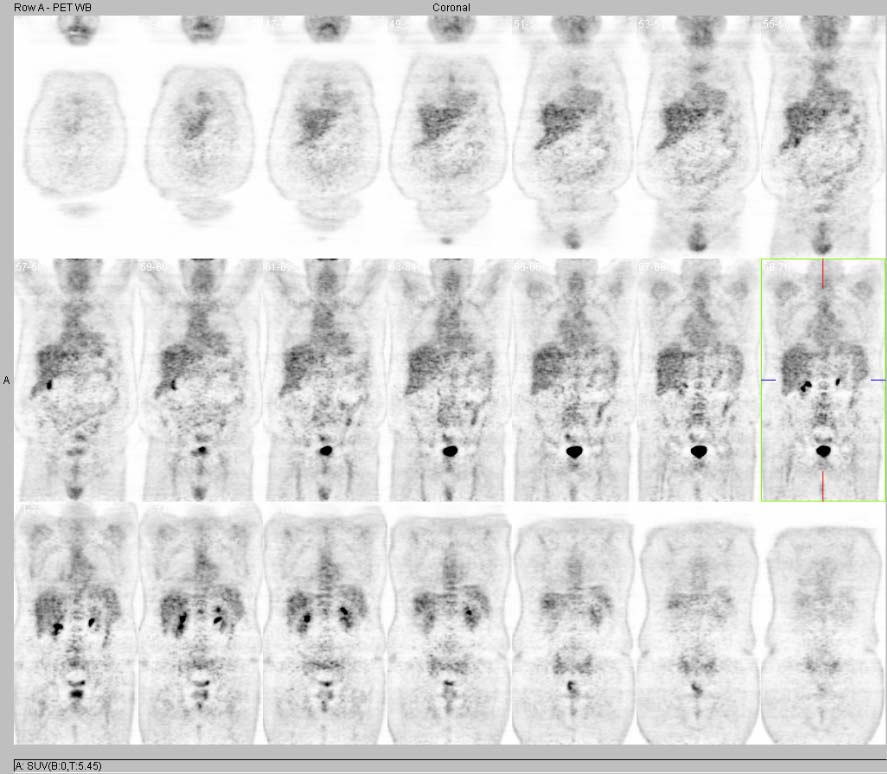
Coronal PET images. View MIP cine in AVI format.
View main image(pt) in a separate image viewer
View second image(pt).
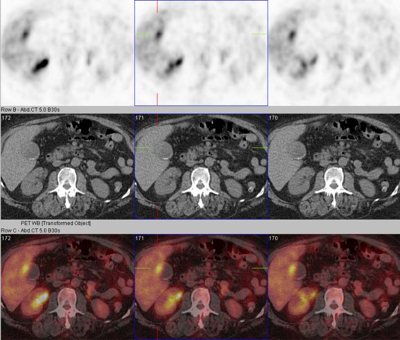
Axial PET/CT images
View third image(ct).
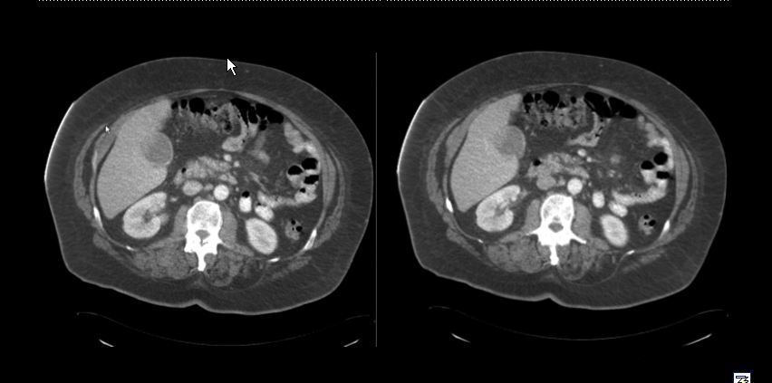
Axial CT
Full history/Diagnosis is available below
Diagnosis: Gallbladder Carcinoma
Full history:
67 year-old man presented with right back pain. Work-up demonstrated a right pleural effusion. The patient subsequently underwent thoracentesis. Cytology was indeterminant.
Radiopharmaceutical:
15.0 mCi F-18 Fluorodeoxyglucose i.v.
Findings:
There is markedly increased FDG uptake in the lateral aspect of the gallbladder, corresponding to a focal area of thickening and nodularity involving the gallbladder wall, which appears to extend into the lumen of the gallbladder and involves the nondependent portion. There are no other regions of abnormally increased FDG uptake within the liver. Cholelithiasis is also noted.
Discussion:
Biliary tract tumors (cholangiocarcinoma, gallbladder carcinoma) are often unresectable at the time of diagnosis. Biliary cancer is the fifth most common GI malignancy. Gallbaldder carcinoma is associated with cholelithiasis, inflammatory bowel disease, porcelain gallbladder, familial polyposis and chronic cholecystitis. Radiographic features include intraluminal soft tissue density, asymmetrically thickened gallbladder wall, direct invasion of the liver, gastrohepatic and hepatoduodenal ligaments and lymph nodes.
The majority of these patients are evaluated after the incidental finding of gallbladder carcinoma in cholecystectomy specimens. FDG-PET is considered to be useful for evaluating malignant gallbladder tumors, especially small tumors, which are difficult to differentiate as benign or malignant using other modalities.
Reference
Christopher D. Anderson M.D., Michael H. Rice M.D., C. Wright Pinson M.D., William C. Chapman M.D., Ravi S. Chari M.D. and Dominique Delbeke M.D., Ph.D. Fluorodeoxyglucose PET imaging in the evaluation of gallbladder carcinoma and cholangiocarcinoma. Journal of Gastrointestinal Surgery, Volume 8, Issue 1, January 2004: 90-97
Koh T, Taniguchi H, Yamaguchi A, Kunishima S, Yamagishi H. Differential diagnosis of gallbladder cancer using positron emission tomography with fluorine-18-labeled fluoro-deoxyglucose (FDG-PET). J Surg Oncol. 2003 Oct; 84(2):74-81.
Koh T, Taniguchi H, Kunishima S, Yamagishi H. Possibility of Differential Diagnosis of Small Polypoid Lesions in the Gallbladder Using FDG-PET. Clin Positron Imaging. 2000 Sep; 3(5):213-218.
Followup:
The patient underwent diagnostic laparoscopy and radical cholecystectomy with central liver wedge resection of segments 4 and 5. Pathology demonstrated gallbladder carcinoma without locoregioal invasion or metastatic lymph nodes.
Major teaching point(s):
Focal increased FDG uptake within the gallbladder should raise concern for early-stage gallbladder carcinoma.
ACR Codes and Keywords:
References and General Discussion of PET Tumor Imaging Studies (Anatomic field:Gasterointestinal System, Category:Neoplasm, Neoplastic-like condition)
Search for similar cases.
Edit this case
Add comments about this case
Return to the Teaching File home page.
Case number: pt142
Copyright by Wash U MO