Case Author(s): Rusty Roberts, M.D., Delphine Chen, M.D. and Henry Royal, M.D. , 6/17/05 . Rating: #D2, #Q4
Diagnosis: Pulmonary angiosarcoma
Brief history:
35 year old cachectic gentleman with chronic dyspnea presents for pulmonary embolism workup.
Images:

Ventilation and perfusion images
View main image(vq) in a separate image viewer
View second image(xr).
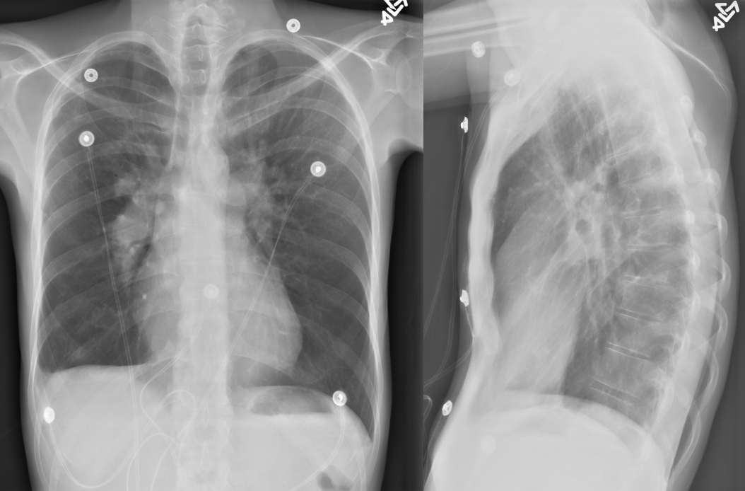
Chest radiographs
View third image(pt).
FDG PET coronal images. View MIP cine in AVI format.
View fourth image(pt).
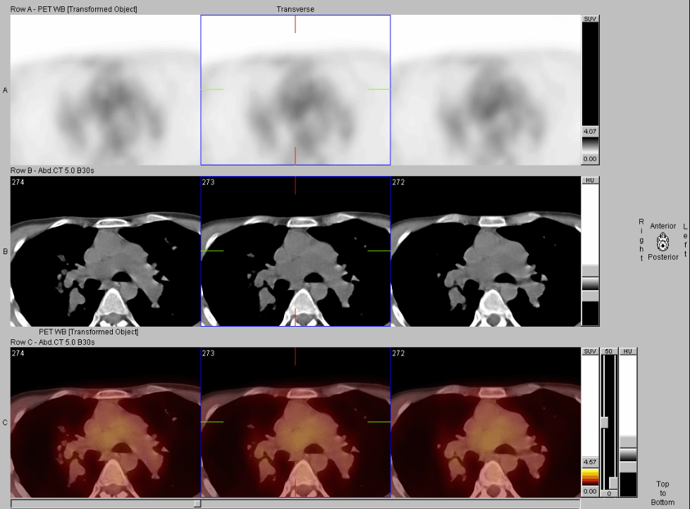
FDG PET/CT images
Full history/Diagnosis is available below
Diagnosis: Pulmonary angiosarcoma
Full history:
35 year old cachectic gentleman with chronic dyspnea presented from an outside institution with a vascular tumor or pulmonary embolus.
Radiopharmaceutical:
29 mCi Xe-133 gas by inhalation and 4.1 mCi Tc-99m MAA i.v.
14.9 mCi F-18 Fluorodeoxyglucose i.v.
Findings:
V/Q: The Xe-133 ventilation images show a uniform distribution of activity on single-breath and washin images in the left lung with mildly decreased ventilation to the right lung with retention of the Xe-133 in the midportion of the right lung. There is absent perfusion to the entire right lung and decreased perfusion to the medial basal and superior segments of the left lower lobe as well as the lingula.
Chest xray: There is marked enlargement of bilateral pulmonary arteries consistent with pulmonary hypertension. There is bilateral pleural parenchymal thickening and scarring. There is a small right pleural effusion noted. The patient has a pectus excavatum deformity of the chest.
FDG PET: Review of the PET images demonstrates no significant areas of FDG uptake within the pulmonary arteries or within the lungs. There is increased FDG uptake within the right ventricle consistent with right ventricular hypertrophy in the setting of longstanding pulmonary stenosis likely secondary to the vascular obstruction by the lesion. No other areas of abnormal FDG uptake are seen on the scan.
Discussion:
Because of the patient's young age and history, a tumor was suspected throughout the workup, however, the imaging tools did not definitively diagnose this patient until the follow up MR demonstrating heterogeneous delayed enhancement. Only then did the clinicians feel confident in proceeding with a endovascular biopsy.
Followup:
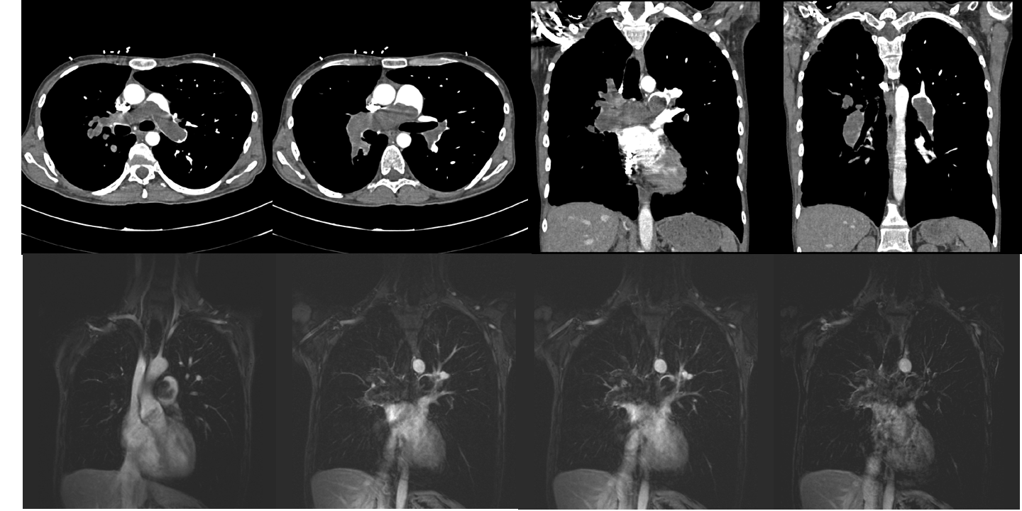
PE protocol CT was then acquired in an attempt to see enhancement of the intravascular lesion. No definitive diagnosis could be made at that time. MRI was then performed. There was heterogeneous enhancement of the pulmonary artery mass. Endovascular biopsy revealed a high grade angiosarcoma of intimal origins.
View followup image(mr).
Collage of CT and MR (pre and post VIBE sequences with the last image having subtraction applied) images
Major teaching point(s):
History and physical are critical to proper interpretation of nuclear and radiologic images.
Differential Diagnosis List
Unilateral perfusion abnormality on VQ: pulmonary vascular mass, pulmonary embolus, congential stenosis, vasculitis, or fibrosing mediastinitis
PET with low metabolic activity: bland thrombus or low metabolic activity within tumor
ACR Codes and Keywords:
References and General Discussion of PET Tumor Imaging Studies (Anatomic field:Lung, Mediastinum, and Pleura, Category:Neoplasm, Neoplastic-like condition)
Search for similar cases.
Edit this case
Add comments about this case
Return to the Teaching File home page.
Case number: pt137
Copyright by Wash U MO