
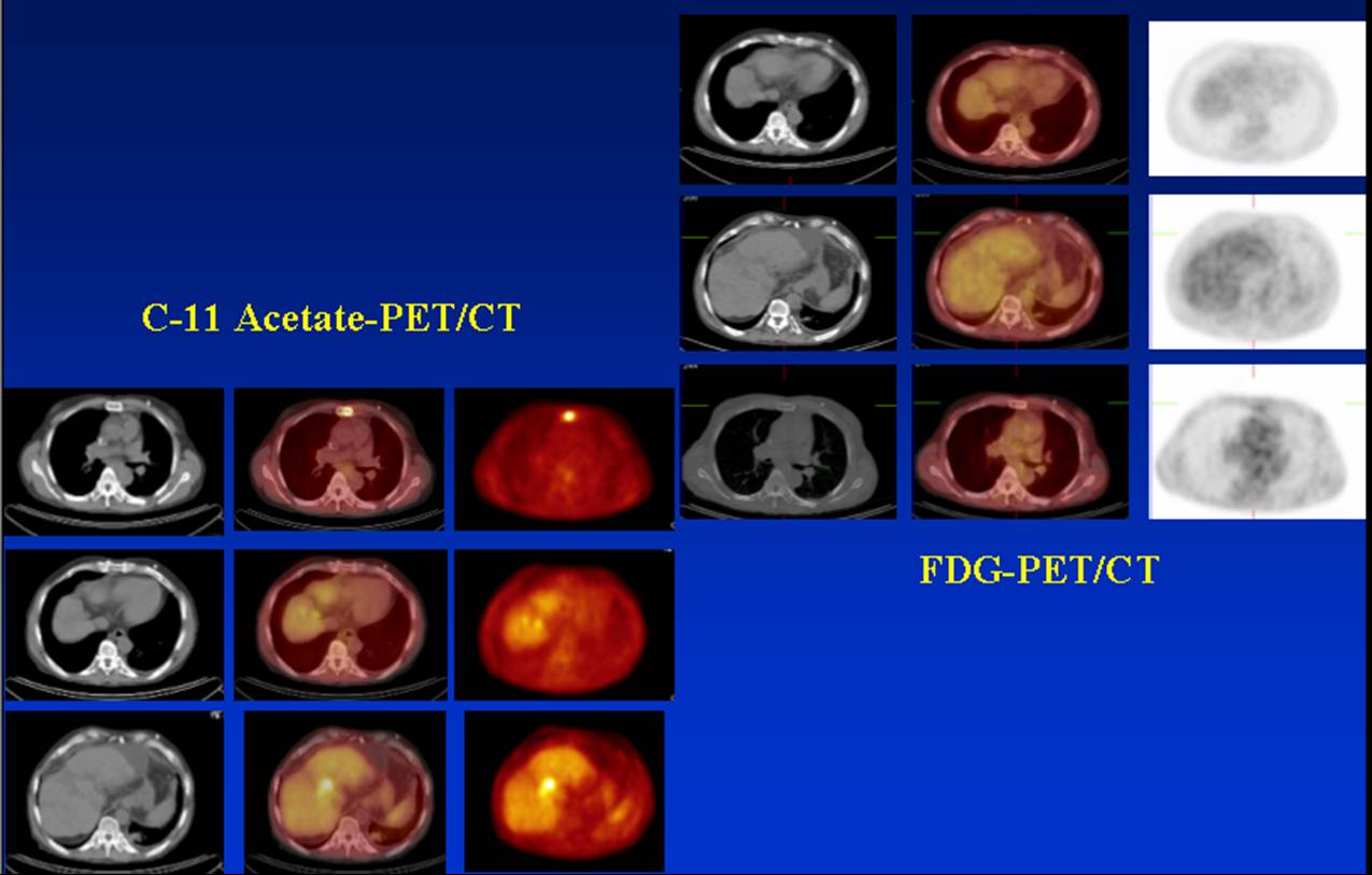
Acetate PET Image.
View main image(pt) in a separate image viewer
View second image(pt). FDG PET image.
View third image(pt). Comparison of fused acetate-PET and FDG-PET.
View fourth image(mr). Axial MRI of the liver.
Full history/Diagnosis is available below
There are several foci of increased uptake in the supraclavicular regions (left greater than right), the left paratracheal, the prevascular (substernal), and bilateral external iliac (left greater than right). These areas are suspicious for lymph node metastases. There is intense C-11 acetate uptake in the upper body of the sternum. There are no corresponding lesions on the bone scintigraphy or the bone windows of the CT examination. There is a small area of increased uptake involving the vertebral body of T1.
No abnormality is seen on FDG-PET to suggest a high-grade hepatocellular carcinoma in the liver. There is no regional or distant metastatic disease.
MRI study demonstrated a 9 mm lesion in segment 8 of the liver with arterial phase enhancement, likely representing hepatocellular carcinoma. At the same level more medially, there is a 1.5 x 1.2 cm lesion and additionally, a 2.1 x 1.9 cm round lesion located within segment 4A centrally, and both of these lesions show borderline arterial phase enhancement which may may represent hepatocellular carcinoma.
Approximately one third to one half of HCCs do not accumulate FDG and will provide false-negative FDG-PET imaging. 11C-Acetate is a metabolic substrate of ß-oxidation and precursors of amino acid and sterol and has proven to be useful in detecting various malignancies; although the sensitivity does not appear as high as with FDG, it may play a complementary role for tumors that are not FDG avid. The study of Ho et al. demonstrated that the poorly differentiated HCCs were detected by FDG-PET and the well-differentiated types were detected by 11C-acetate-PET. It is also interesting to observe that approximately 30% of the HCCs were both FDG and 11C-acetate avid and actually demonstrated heterogeneity of metabolism in different parts of the same tumor. The authors concluded that the dual-isotope technique can be very useful to evaluate indeterminate hepatic lesions. When the lesion accumulates both tracers or accumulates only 11C-acetate, HCC is high in the differential diagnosis; when it accumulates only FDG, a non-HCC malignancy should be considered; and if it is negative for both tracers, a benign pathology is more likely.
HCC has long been a disease with few therapeutic options, but recently the results of liver transplantation have been very favorable when the disease is detected early enough. Transplant results in patients with early-stage HCC have been so positive that the United Organ Sharing Network (UNOS) has changed its allocation procedures to give these patients higher priority, allowing them to be transplanted quickly. According to recent studies, when patients with hepatitis-C and stage I or stage II HCC are transplanted, their five-year survival is around 70 percent about the same as for patients with hepatitis-C who do not have cancer.
References:
1. FOCAL SPOT, FALL/WINTER 2003/2004.
2. Ho CL, Yu, SCH, Yeung DWC. 11C-Acetate PET Imaging in Hepatocellular Carcinoma and Other Liver Masses. J. Nucl. Med. 2003; 44(2): 213-221.
3. Delbeke D and Pinson CW. 11C-Acetate: A New Tracer for the valuation of Hepatocellular Carcinoma. J. Nucl. Med. 2003; 44(2): 222 - 223.
References and General Discussion of PET Tumor Imaging Studies (Anatomic field:Gasterointestinal System, Category:Neoplasm, Neoplastic-like condition)
Return to the Teaching File home page.