FDG-PET whole body. View MIP cine in AVI format.
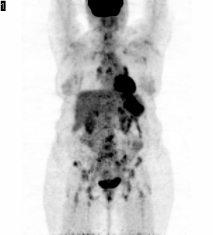
View main image(pt) in a separate image viewer
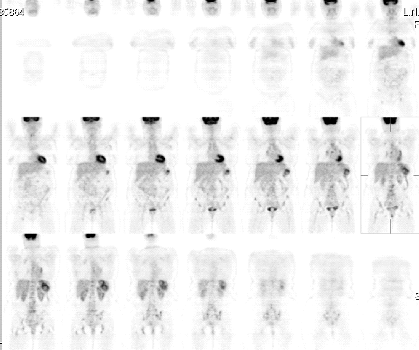
View second image(pt). FDG-PET coronal image
View third image(pt). Fused image of PET and CT
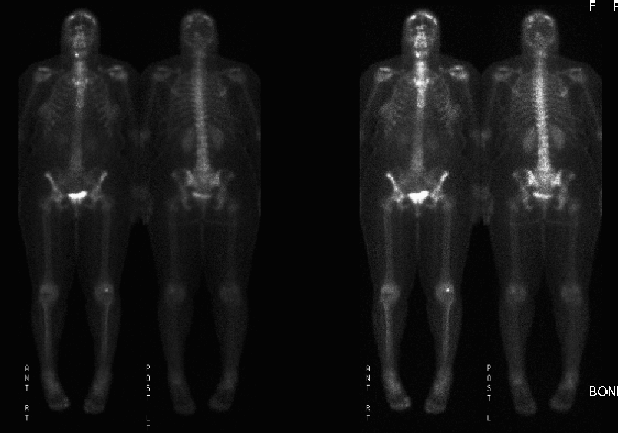
View fourth image(bs). Whole body bone scintigraphy
Full history/Diagnosis is available below
Sarcoidosis is a disease that causes inflammation of the bodyĺs tissues. In sarcoidosis, the inflammation produces nodules or granulomas in the tissues. The inflammation of sarcoidosis can occur in almost any organ and always affects more than one. Most often, the inflammation starts in either the lungs or the lymph nodes.
Sarcoidosis occurs most commonly in adults between the ages of 20 and 40. The cause of sarcoidosis is not known; This diagnosis is what is known as a "diagnosis of exclusion". In general, if a person has moderate to severe symptoms or treatment is going to be given, tissue diagnosis is recommended.
Most people with sarcoidosis have no symptoms. Some have only one symptom, while still others have many. Symptoms typically depend on which organs the disease affects. In most patients, the inflammation that causes the granulomas gets better with or without treatment and the lumps go away. In others, however, the lumps do not heal or disappear, and the tissues remain inflamed. If untreated, these tissues can become scarred.
The treatment of sarcoidosis depends on a personĺs symptoms. 60% patients with sarcoidosis do not need treatment. But, for some patients, intense treatment is required, especially if there is critical organ involvement. Treatment is done to control symptoms or to improve the function of organs affected by the disease.
Sarcoidosis granulomas result from a response of the immune system. Thus, most medications used to treat sarcoidosis suppress the immune system. Treatment may or may not affect the long-term outcome of the disease.
Reference: Web site: U.S. Department of Health and Human Services.
References and General Discussion of PET Tumor Imaging Studies (Anatomic field:Genitourinary System, Category:Inflammation,Infection)
Return to the Teaching File home page.