Case Author(s): Rusty Roberts M.D. and Barry A. Siegel M.D. , . Rating: #D3, #Q4
Diagnosis: Hepatic metastasis from colon cancer and incidental sarcoidosis
Brief history:
65-year-old man presents for staging of rectosigmoid adenocarcinoma. The primary tumor was resected approximately 5 weeks before PET imaging. No radiation or chemotherapy has been administered.
Images:

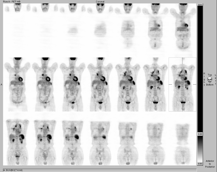
Coronal PET images. A
cine of the MIP images is also available
View main image(pt) in a separate image viewer
View second image(pt).
Axial PET images of the abdomen
View third image(pt).
PET/CT fusion images of the upper abdomen
View fourth image(pt).
PET/CT fusion images of the upper lobes
Full history/Diagnosis is available below
Diagnosis: Hepatic metastasis from colon cancer and incidental sarcoidosis
Full history:
65-year-old man presented for staging of rectosigmoid adenocarcinoma. The primary tumor was resected approximately 5 weeks before PET. He also has a history of lymphoma 23 years ago (site unknown), which was surgically resected and treated with radiation and chemotherapy. He currently has no known lymphoma recurrence. Additionally, he has findings of sarcoidosis on a CT examination 3 months ago, with a past lung biopsy showing granulomatous inflammation.
Radiopharmaceutical:
14.9 mCi F-18 Fluorodeoxyglucose i.v.
Findings:
Intense FDG uptake is seen within the parahilar lung parenchyma. This corresponds to nodular interstitial thickening and alveolar opacifications within the lungs. Scattered pulmonary nodules, all less than 1 cm in diameter, are seen bilaterally. The FDG uptake within these nodules is highly variable. There are bilateral enlarged hilar lymph nodes which are partially calcified and FDG-avid. The medial segment of the right middle lobe is atelectatic and has intense FDG uptake. The spleen is also intensely FDG-avid. All of the above findings can be ascribed to the patient's presumed sarcoidosis.
There are four discrete FDG-avid lesions within the liver, which are suspicious for metastases. The largest of these lesions is in the posterior segment of the right lobe, and this has a low-attenuation CT correlate. Two smaller FDG-avid foci are seen within the dome of the liver. One of these has the appearance of being within the lung secondary to respiratory-motion misregistration between the PET data and the CT data used for attenuation correction. A fourth lesion is seen within the anterior segment of the right lobe. Only the posterior segment lesion has a CT correlate. FDG uptake elsewhere in the liver is patchy. None of these hepatic lesions were seen on the CT performed 3 months ago. There is mildly increased FDG uptake seen within the midline incision.
Discussion:
The hepatic lesions are consistent with metastatic colon cancer.
Intense FDG uptake within the lungs, mediastinum, and the spleen is likely secondary to the patient's sarcoidosis. Involvement of these sites by the long quiescent lymphoma or the new colon carcinoma are unlikely explanations for these findings.
Lewis PJ, Salama A. Uptake of fluorine-18-fluorodeoxyglucose in sarcoidosis. J Nucl Med 1994; 35:1647-1649.
Milman N, Mortensen J, Sloth C. Fluorodeoxyglucose PET scan in pulmonary sarcoidosis during treatment with inhaled and oral corticosteroids. Respiration 2003; 70:408-413.
Warshauer DM, Lee JKT. Imaging manifestations of abdominal sarcoidosis. AJR 2004; 182:15-28.
Major teaching point(s):
Violation of Occam's razor (one should not increase, beyond what is necessary, the number of entities required to explain anything) is necessary in this case. A good patient history is vital to the correct interpretation of PET images. Active granulomatous disease is among the most common causes of abnormal FDG uptake that can be confused with cancer.
Differential Diagnosis List
Metastatic disease, lymphoma, sarcoidosis, or infectious disease.
ACR Codes and Keywords:
References and General Discussion of PET Tumor Imaging Studies (Anatomic field:Gasterointestinal System, Category:Neoplasm, Neoplastic-like condition)
Search for similar cases.
Edit this case
Add comments about this case
Return to the Teaching File home page.
Case number: pt129
Copyright by Wash U MO

