Case Author(s): Eric Hutchins, MD and Keith Fischer, MD M.D. , 01/28/05 . Rating: #D3, #Q4
Diagnosis: Normal Variant - Hypermetabolic Fat in the Interatrial Septum
Brief history:
79 year old female with a large palpable right breast mass. Biopsy yielded a diagnosis of invasive ductal carcinoma.
Images:
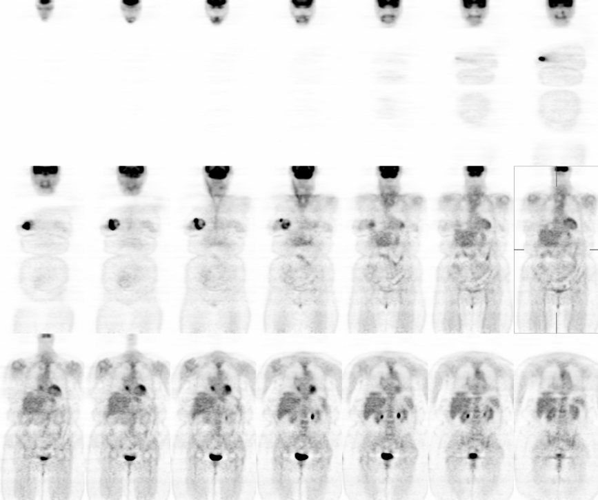
Coronal PET Images
View main image(pt) in a separate image viewer
View second image(pt).
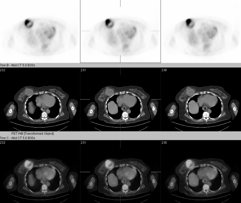
Selected Axial PET/CT Images
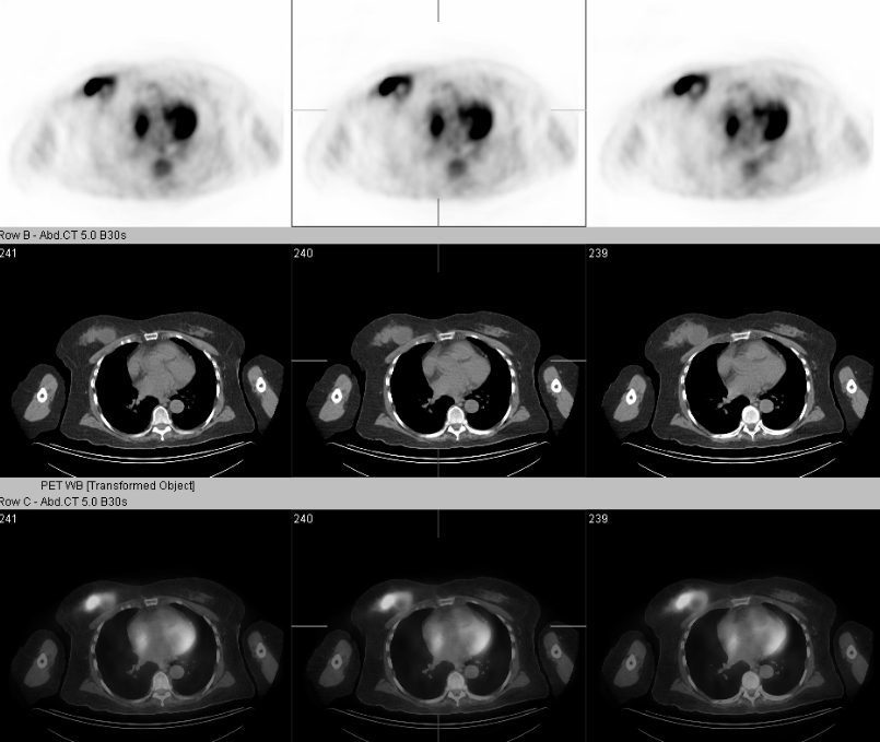
View third image(pt).
Selected Axial PET/CT Images
Full history/Diagnosis is available below
Diagnosis: Normal Variant - Hypermetabolic Fat in the Interatrial Septum
Full history:
79 year old female with a palpable right breast mass. Ultrasound guided biopsy performed 9 days prior to PET/CT imaging yeilded a diagnosis of invasive ductal carcinoma.
Radiopharmaceutical:
F-18 Fluorodeoxyglucose
Findings:
Coronal images demonstrate the hypermetabolic right breast mass. There is no evidence of hypermetabolism to suggest right axillary or internal mammary nodal metastases. There is a focus of hypermetabolism in the mediastinum of uncertain significance. No other hypermetabolic foci are present to suggest distant metastatic disease.
Selected images through the right breast mass demonstrate heterogeneous hypermetabolism.
Selected images through the heart show the above-mentioned focus of hypermetabolism to be within fat of the interatrial septum.
Discussion:
Hypermetabolic brown fat within the interatrial septum is a normal variant and should not be reported as a metastasis. There are also fat-containing tumors of the interatrial septum including rhabdomyoma, rhabdomyosarcoma, myxoma, lipoma, and liposarcoma. A common reason for an enlarged, fat-containing septum is lipomatous hypertrophy of the interatrial septum which can also be hypermetabolic on PET imaging. If hypermetabolism is seen in the region of the right heart, correlation with CT images is necessary for further evaluation.
Reference: Fan CM, et al., "Lipomatous Hypertrophy of the Interatrial Septum: Increased Uptake on FDG-PET" AJR Jan 2005;184:339-342.
Followup:
At surgery, the right breast tumor was 7.5 cm in size and 1 of 14 right axillary lymph nodes was positive for metastases. The size of the nodal metastatic implant was 1mm. This illustrates the low sensitivity of FDG-PET for nodal micrometastaes.
Major teaching point(s):
If increased FDG uptake is seen in right heart region, correlate with CT or MRI.
ACR Codes and Keywords:
References and General Discussion of PET Tumor Imaging Studies (Anatomic field:Heart and Great Vessels, Category:Normal, Technique, Congenital Anomaly)
Search for similar cases.
Edit this case
Add comments about this case
Return to the Teaching File home page.
Case number: pt127
Copyright by Wash U MO