Case Author(s): Rusty Roberts M.D. and Henry Royal M.D. , . Rating: #D3, #Q3
Diagnosis: Hodgkin's disease
Brief history:
17-year-old with stage IV-B Hodgkin's disease. 7 months previously, presented for restaging and evaluation of treatment post ABVD regimen (Adriamycin, bleomycin, vinblastine, dacarbazine) times six cycles. She had several one-week delays secondary to neutropenia. She finished her chemotherapy approximately 2 weeks prior to the first image.
Images:
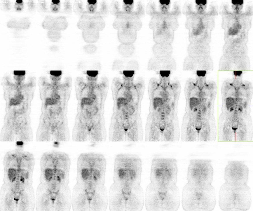
Coronal FDG PET images of the most recent exam.
It may be useful to view the last 3 exams in sequence in AVI format.
View main image(pt) in a separate image viewer
View second image(pt).
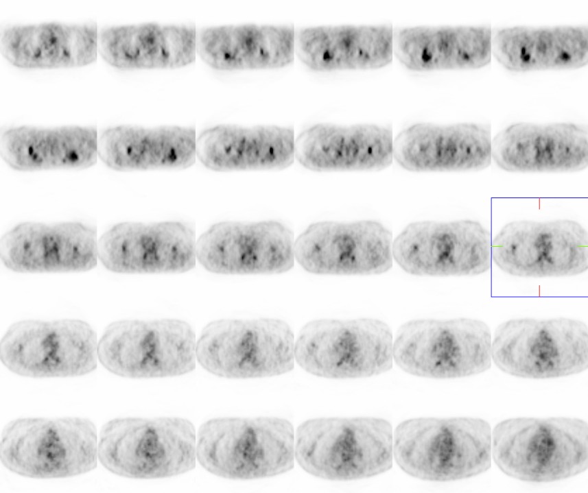
Axial FDG PET images of current study.
View third image(pt).
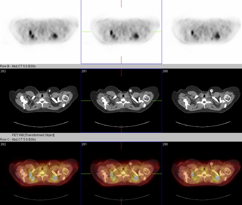
Axial fused PET/CT images of current study.
View fourth image(pt).
Axial fused PET/CT images of current study.
Full history/Diagnosis is available below
Diagnosis: Hodgkin's disease
Full history:
She was a 17-year-old who was diagnosed with stage IV-B Hodgkin's disease 7 months prior to the first rotating MIP images. She received chemotherapy with an ABVDregimen (Adriamycin, bleomycin, vinblastine, dacarbazine) times six cycles. She had several one-week delays secondary to neutropenia. She finished her chemotherapy approximately 2 weeks prior to the last rotating MIP image which correspond to the coronal, axial and fused static images.
Radiopharmaceutical:
12.6 mCi F-18 Fluorodeoxyglucose i.v.
Findings:
The initial MIP image demostrates extensive nodal disease within the neck, thorax and abdomen. This was stage IV-B Hodgkin's disease at presentation.
The second MIP image, 5 months later, demonstrates resolution of her disease.
The third MIP image reveals intense FDG activity within the neck and upper chest. Subsequent fused PET/CT imagery better defines the activity to fat not muscle. This is consistent with brown fat activity and not recurrence.
Discussion:
The difficulty interpreting PET images alone should not be underestimated. Without an accompanying CT images acquired near the same time, this patient might have been misinterpreted as a Hodgkin's disease recurrence. The other misinterpretation, which is common without fused CT imagery, was muscle activity. The actual test was a PET/CT allowing direct comparison.
Brown fat is necessary for thermoregulation and begins activating prior to muscular contractions, ie shivering. Valium or other short acting benzodiazepines have been successfully used to counteract both the brown fat and muscular activity in PET imaging.
The exact mechanisms have as yet to be well defined but both cold temperatures and anxiety are know causes of increased activity. Anxiety originated FDG activity is probably triggered by the increased adrenergic nervous system within brown fat. An additional point to ponder concerns the time of year and temperature of the department when the study was performed. In colder months, when the last PET in this case was acquired, one expects more brown fat activity.
Followup:
"Focal FDG Uptake in Mediastinal Brown Fat Mimicking Malignancy: A Potential Pitfall Resolved on PET/CT." Truong, M. T., et al. AJR 2004; 183:1127-1132. http://www.ajronline.org/cgi/content/figsonly/183/4/1127
Differential Diagnosis List
Hodgkin's recurrence, brown fat, muscle activity
ACR Codes and Keywords:
References and General Discussion of PET Tumor Imaging Studies (Anatomic field:Vascular and Lymphatic Systems, Category:Neoplasm, Neoplastic-like condition)
Search for similar cases.
Edit this case
Add comments about this case
Return to the Teaching File home page.
Case number: pt125
Copyright by Wash U MO