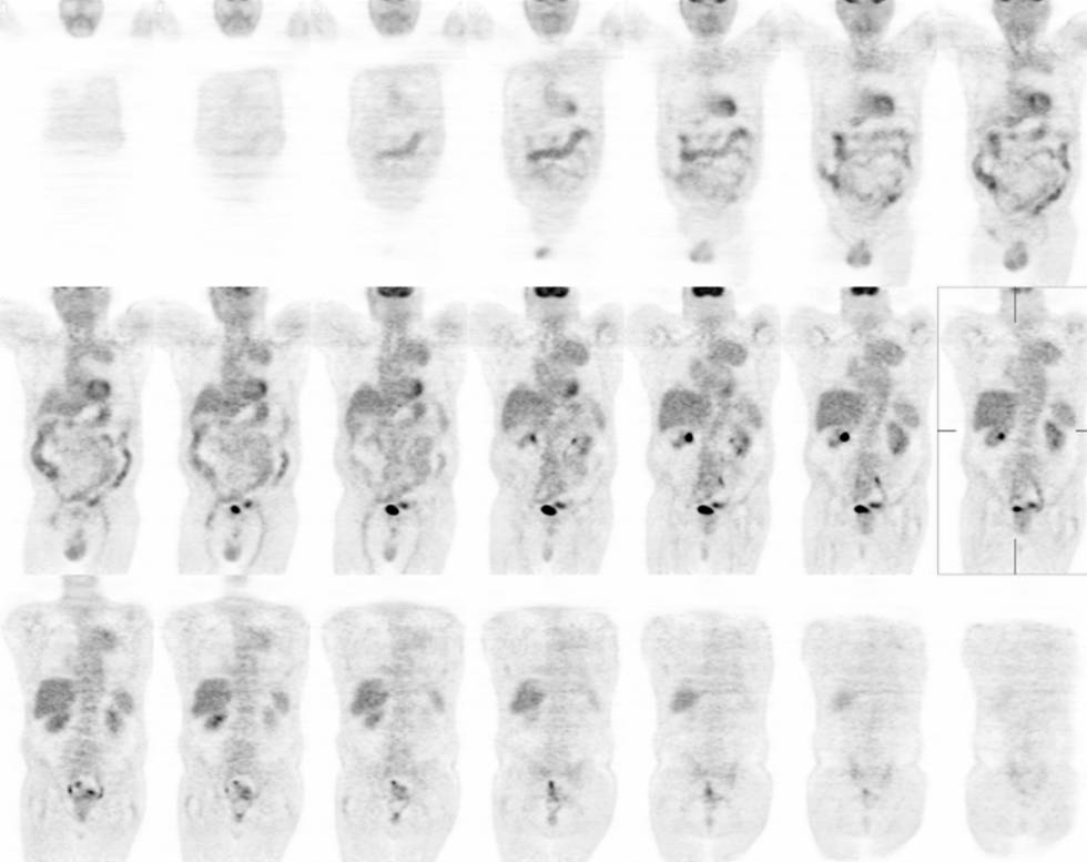
Coronal images from a FDG-PET examination.
View main image(pt) in a separate image viewer
View second image(pt). Selected axial images from a FDG-PET examination.
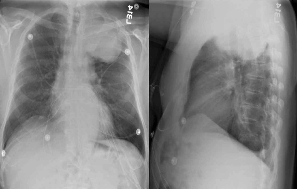
View third image(xr). PA and Lateral Chest Radiographs.
Full history/Diagnosis is available below
Selected axial images through the tumor show the mass to be contiguous with the aorta at the level of the aortic arch. There are several coarse calcifications with in the mass.
Chest radiographs demonstrate a left apical mass with a sharp distinction between the lesion and normal lung. Also note the indistinct margin and obtuse angle between the lesion and the mediastinum/chest wall.
When evaluating a chest lesion on chest radiographs, the angle between the lesion and the chest wall/mediastinum can be helpful in localizing the lesion. If the lesion is extrapulmonary (i.e. pleural) then it will often form an obtuse angle with the chest wall/mediastinum.
Solitary fibrous tumor of the pleura originates from submesothelial connective tissue of the pleura. Majority (80%) arise from the visceral plerua. In a retrospective review of 360 cases, 88% were benign. Average size was 6 cm, with a range of 1-36 cm. Pathology was not able to predict prognosis (local recurrence) with certainty. Thus, these tumors were surgically removed (Briselli et al.).
Recently, a retrospective review of 32 patients with pleural thickening on CT who underwent FDG-PET showed that malignant fibrous tumors have metabolism similar to that of benign processes and caused false-negative results for malignancy (Kramer et al.).
References:
Briselli M, Mark EJ, Dickersin GR. Solitary fibrous tumors of the pleura: eight new cases and review of 360 cases in the literature. Cancer. 1981 Jun 1;47(11):2678-89.
Kramer H, Pieterman RM, Slebos DJ, Timens W, Vaalburg W, Koeter GH, Groen HJ. PET for the evaluation of pleural thickening observed on CT. J Nucl Med. 2004 Jun;45(6):995-8.
References and General Discussion of PET Tumor Imaging Studies (Anatomic field:Lung, Mediastinum, and Pleura, Category:Neoplasm, Neoplastic-like condition)
Return to the Teaching File home page.