Case Author(s): Rusty Roberts M.D. and Barry A. Siegel M.D. , 10/29/04 . Rating: #D3, #Q4
Diagnosis: Pseudomembranous Colitis
Brief history:
75-year-old man with a history of squamous cell carcinoma of the mouth, which was treated with wide local excision followed by radiation therapy. The patient had positive margins after his surgery. We were asked to restage the patient's tumor nine months later while he was hospitalized for dehydration and percutaneous endoscopic gastrostomy (PEG) placement.
Images:
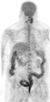
Anterior MIP FDG-PET image. View cine of this image as an avi file
View main image(pt) in a separate image viewer
View second image(pc).
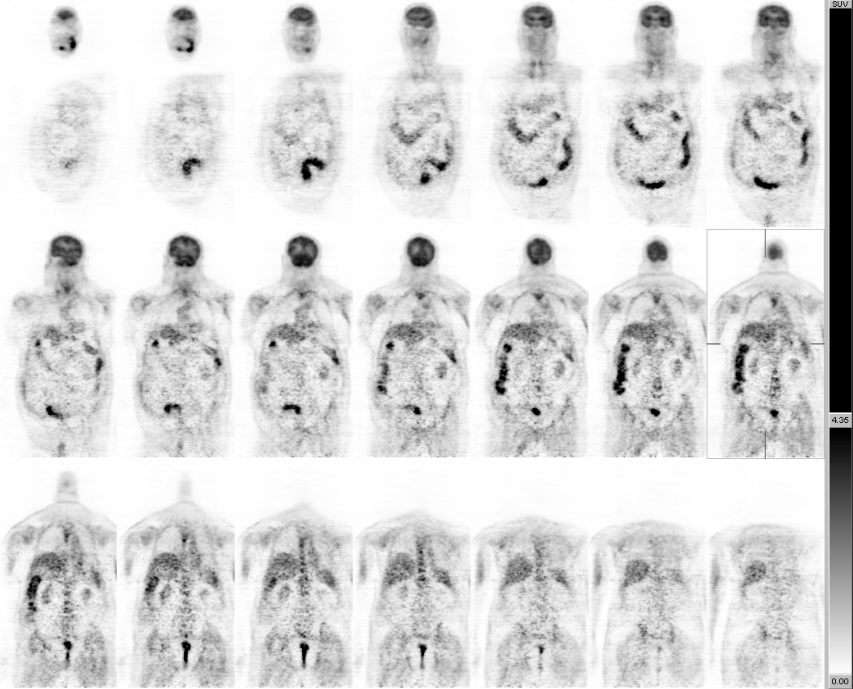
Coronal FDG-PET images
View third image(pt).
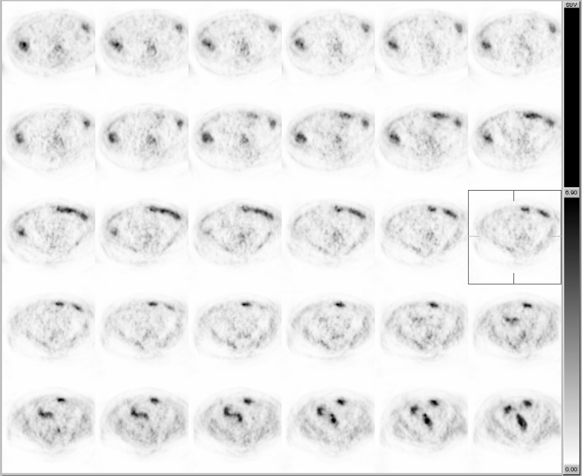
Axial FDG-PET images through the upper abdomen
View fourth image(ct).
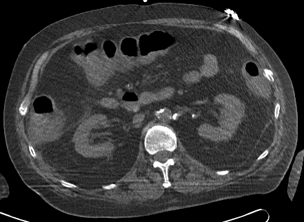
Axial CT through the upper abdomen
Full history/Diagnosis is available below
Diagnosis: Pseudomembranous Colitis
Full history:
75-year-old man with a history of squamous cell carcinoma of the mouth, which was treated with wide local excision of the mouth, left lip, and cheek, followed by radiation therapy. The patient had positive margins after his surgery. He presented to the ENT clinic with pain, weight loss, dysphagia, and diarrhea. The diarrhea began one week before admission, after the placement of a nasogastric tube for Jevity feedings. He was admitted from the clinic for dehydration and percutaneous endoscopic gastrostomy (PEG) placement.
The PEG was placed and an FDG-PET/CT study was ordered for restaging because an oral biopsy was positive for recurrent squamous cell carcinoma. One month earlier a conventional FDG-PET study had been equivical for distant disease.
Radiopharmaceutical:
14.9 mCi F-18 Fluorodeoxyglucose i.v.
Findings:
Intensely increased FDG uptake is seen within areas of soft tissue thickening in the neck noted on CT (not shown). There is an inferior extension of this FDG-avid soft tissue thickening along the anterior aspect of the left mandibular body. A large FDG-avid left submandibular lymph node also is seen near the angle of the mandible. There is a focal area of increased FDG uptake in the floor of the mouth. No discrete mass was seen on the corresponding noncontrast CT images (not shown), however.
The ascending and descending colon demonstrate moderately intense, diffusely increased FDG uptake. There is also diffuse thickening of the ascending and descending colonic walls, particularly involving the rectosigmoid on CT. There is relative sparing of the transverse colon, however. A Foley catheter is present in the urinary bladder.
Discussion:
Abnormally increased FDG uptake may due to tumor and inflammatory conditions. In this patient, the unsuspected finding of diffusely increased colonic uptake was noted. This is not a typical pattern for a primary colonic cancer or for metastatic disease, especially from an oral squamous cell carcinoma. Rather, it suggests a diffuse colitis.
Followup:
These findings prompted a discussion with the referring physician concerning possible infectious etiologies for the colonic FDG uptake and the patient's recent diarrhea. A stool sample was positive for Clostridium difficile, and treatment was initiated.
Major teaching point(s):
Uptake of FDG in the esophagus, stomach, small intestine, and colon can be seen as a highly variable, but normal feature on PET images. The tracer is localized in the wall of the visualized gastrointestinal tract structure, rather than within the lumen. No effective means for eliminating gastrointestinal tract activity have been devised, but experienced interpreters can distinguish this normal variant from disease in most cases.
The finding of focal intense FDG uptake in the colon should raise the question of a primary colonic neoplasm. The finding of a long segment of intense FDG uptake should raise the question of colitis. Correlation with the patient's history and available imaging studies should be performed. Depending on the clinical circumstances, recommendation of additional imaging or colonoscopy may be appropriate.
References
1. Tatlidil R, Jadvar H, Bading JR, Conti PS. Incidental colonic fluorodeoxyglucose uptake: correlation with colonoscopic and histopathologic findings. Radiology 2002; 224:783-7.
2. Hannah A, Scott AM, Akhurst T, Berlangieri S, Bishop J, McKay WJ.
Abnormal colonic accumulation of fluorine-18-FDG in pseudomembranous colitis. J Nucl Med 1996; 37:1683-5.
Differential Diagnosis List
Prominent physiologic (normal) colonic uptake of FDG
Inflammatory bowel disease ()e.g., ulcerative or granulomatous colitis)
Infectious colitis
Attenuation-correction artifact (on PET/CT) caused by contrast agent in the bowel
ACR Codes and Keywords:
References and General Discussion of PET Tumor Imaging Studies (Anatomic field:Gasterointestinal System, Category:Inflammation,Infection)
Search for similar cases.
Edit this case
Add comments about this case
Return to the Teaching File home page.
Case number: pt122
Copyright by Wash U MO