Case Author(s): Akash Sharma, M.D. and Jerold W. Wallis, M.D. , 09/21/2004 . Rating: #D3, #Q5
Diagnosis: Nodular fatty infiltration in the liver
Brief history:
42 year old woman underwent chemotherapy 10 years ago presents for follow-up evaluation.
Images:
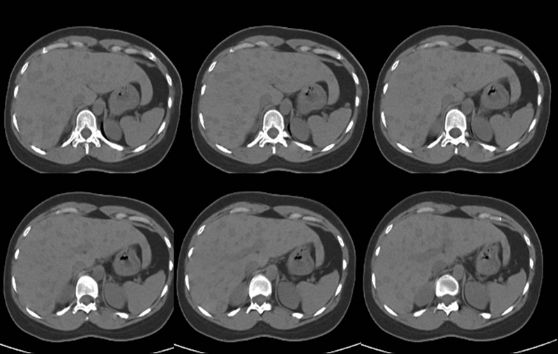
Selected images from the non-contrast CT from patient's PET-CT study
View main image(ct) in a separate image viewer
View second image().
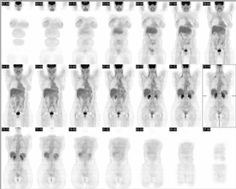
Coronal PET images
View third image(pt).
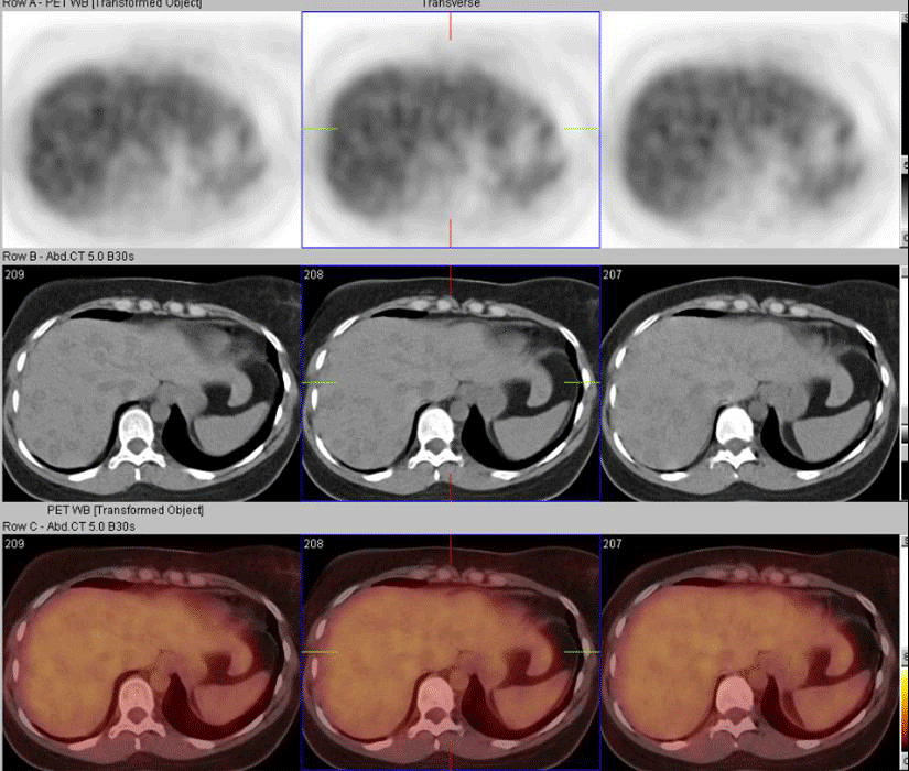
Selected fused PET-CT images
View fourth image(pt).
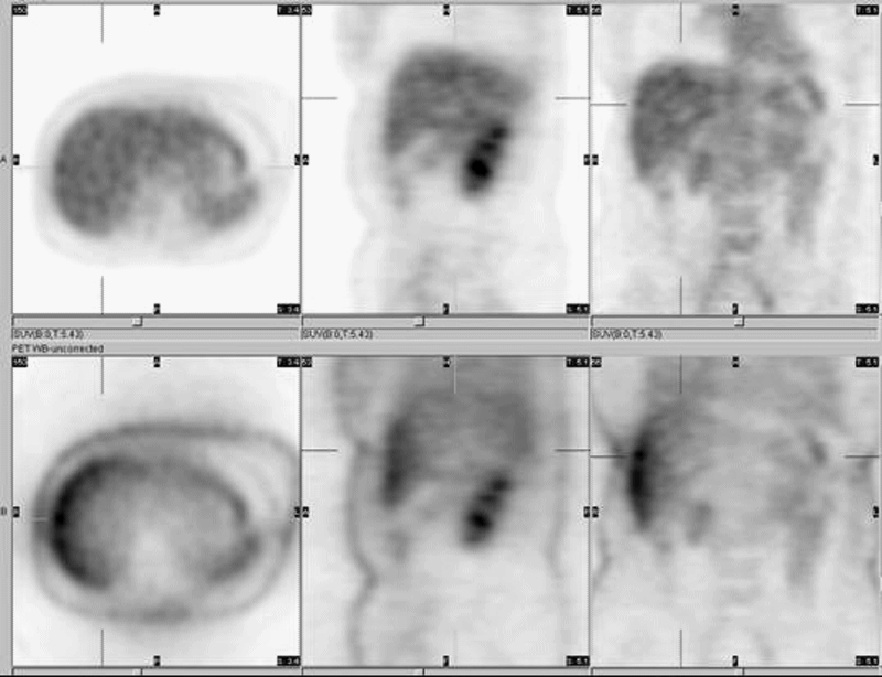
Selected attenuation corrected and non-attenuation corrected images
Full history/Diagnosis is available below
Diagnosis: Nodular fatty infiltration in the liver
Full history:
42 year old woman who had lymphoma and underwent chemotherapy 10 years ago. She has had subsequent annual follow-ups with gallium scintigraphy and also had a PET scan in March 2003. The prior PET scan was performed at Mallinckrodt Institute of Radiology and demonstrated mild uptake in the central mesenteric region of the abdomen, which was interpreted as abnormal. Current PET-CT is for follow-up evaluation for recurrent metastatic disease.
Radiopharmaceutical:
14.1 mCi F-18 Fluorodeoxyglucose i.v.
Findings:
PET-CT:
1. No definite evidence for recurrent disease on FDG imaging. Very mild uptake in central mesenteric area is unchanged and most likely represents post-treatment fibrosis.
2. Multiple low-attenuation targetoid foci in the liver on the CT portion of the study are of unknown clinical significance, and have progressed since the prior exam. While treated lymphoma can look like this (but usually has a greater calcification component), the possibility of low grade recurrent/residual tumor can not be excluded. Given prior history of lymphoma, a contrast enhanced CT or MRI of the adbomen, or liver biopsy, is recommended for further evaluation.
Discussion:
In a patient with known prior lymphoma, multiple liver lesions on the noncontrast CT during PET-CT are not evaluated completely due to the lack of contrast. Given no definite uptake in these lesions (on both attenuation-corrected and non-attenuation corrected images), the differential diagnosis remains wide. Additional imaging such as MRI is need to differentiate benign entities such as fatty infiltration which sometimes can give this appearance on CT.
Followup:
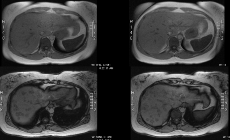
Abdominal MRI:
There are numerous small nodular lesions within the liver. These are not well seen on the T2 inversion recovery images or HASTE images. They demonstrate increased signal intensity on the T2 TURBO spin echo images without fat saturation. They are best seen on the out of phase T1 images. They are not seen on the in-phase T1-weighted images. The lesions do not contrast enhance. They do not exert mass effect or distort adjacent blood vessels. They are felt to be consistent with nodular fatty infiltration.
Based on MRI findings, the lesions were thought to be a nodular pattern of fatty infiltration, atypical but not uncommon. A followup CT evaluation three months later did not show any change in size or enhancement characteristics of these lesions.
View followup image(mr).
Selected MRI images (in and out of phase)
Differential Diagnosis List
Based on PET-CT findings:
1. Low grade recurrence of lymphoma
2. Healed lymphoma (minus calcifications)
3. Fatty infiltration
Based on PET-CT and MRI:
1. Nodular fatty infiltration.
ACR Codes and Keywords:
References and General Discussion of PET Tumor Imaging Studies (Anatomic field:Gasterointestinal System, Category:Metabolic, endocrine, toxic)
Search for similar cases.
Edit this case
Add comments about this case
Return to the Teaching File home page.
Case number: pt120
Copyright by Wash U MO