Case Author(s): Akash Sharma, M.D. and Keith C. Fischer, M.D. , . Rating: #D3, #Q4
Diagnosis: Residual inflammation from post-radiation infection
Brief history:
44-year-old woman with cervical cancer who has completed both radiation therapy and chemotherapy. (View second and third image simultaneously)
Images:

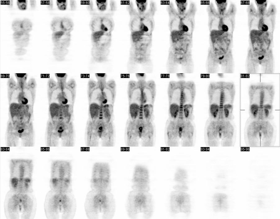
Selected coronal images
View main image(pt) in a separate image viewer
View second image(pt).
Selected fused PET-CT images 01-19-2004
View third image(pt).
Selected fused PET-CT images 07-03-2003
Full history/Diagnosis is available below
Diagnosis: Residual inflammation from post-radiation infection
Full history:
44 year old woman, status post chemotherapy, external beam radiation therapy, and brachytherapy for stage IIB cervical cancer who now has evidence of drainage per vagina approximately three months after treatment completion. There is a possible vesicovaginal fistula based on physical examination. PET-CT performed to restage as part of current workup.
Radiopharmaceutical:
15 mCi F-18 Fluorodeoxyglucose i.v.
Findings:
Current PET-CT:
Compared to the prior examination of 7-3-03, there is again intense abnormal uptake posterior to the bladder; however, there is a central area of decreased activity suggesting necrosis has occurred. No abnormal uptake is seen in the lymph nodes of the pelvis or abdomen. No distant metastatic spread is seen.
Prior PET-CT from 7-2003:
An area of intense uptake of FDG in the cervix corresponding to a large cervical mass, known to be cervical cancer. No other areas of abnormal uptake are identified to suggest metastatic disease.
Discussion:
Given the findings, it is difficult to entirely exclude the possibility fo residual tumor. However, it should be noted that infection can give this appearance. The central necrosis and purulent drainage on physical examination make the likelihood of infection equally feasible. The inability to differentiate tumor from active infection or inflammation is a well-known limitation of PET imaging and clinical information, physical examination findings, and follow-up are critical to provide a more complete evaluation.
Followup:
Patient underwent a dilatation and curettage which revealed significant necrotic and purulent debris with acute and chronic inflammatory changes presumed to be from radiation. No evidence of malignancy was present.
The patient continued to have effective local control of tumor but was found to have mesenteric metastatic disease on a follwup PET-CT evaluation five months later.
View followup image(pt).
Coronal PET images with new mesenteric uptake but no significant uterine uptake 06-02-2004
Differential Diagnosis List
Residual tumor versus acute infection vs. acute or chronic (post-radiation) inflammation
ACR Codes and Keywords:
References and General Discussion of PET Tumor Imaging Studies (Anatomic field:Genitourinary System, Category:Inflammation,Infection)
Search for similar cases.
Edit this case
Add comments about this case
Return to the Teaching File home page.
Case number: pt119
Copyright by Wash U MO

