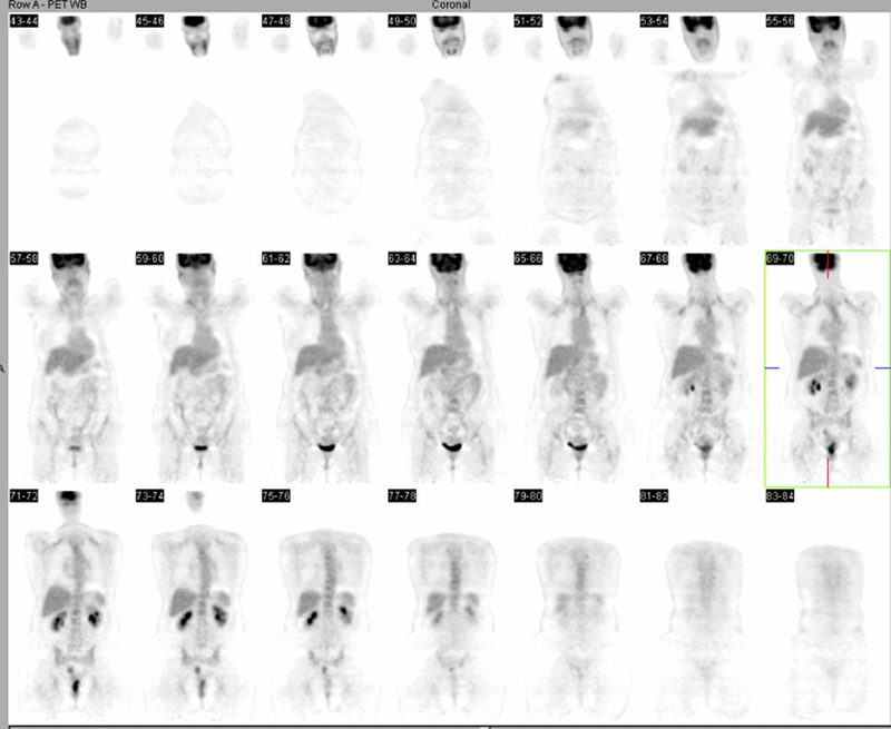

Selected coronal PET images
View main image(pt) in a separate image viewer
View second image(pt). Selected fused PET-CT images
View third image(pt). Non-attenuation-corrected (NAC) and attenuation-corrected (AC) coronal PET images
Full history/Diagnosis is available below
The corresponding CT images in this second region demonstrate barium in the colon, likely a residual from a previous CT study. The apparently increased FDG uptake in this region on the attenuation-corrected (AC) images is secondary to an attenuation-correction artifact. No other foci of abnormal FDG uptake are noted in the abdomen or pelvis. No adenopathy is evident. Incidental mild uptake in the supraclavicular fat and the deep cervical fat at the skull base is noted as a normal variant.
The artifactually increased "activity" in the second focus is not seen on the Non-attenuation-corrected (NAC)images.
In hybrid PET-CT scanners, attenuation correction typically is performed using the CT data. By comparison, in a conventional PET scanner, this is done using the data from a transmission scan performed with a Ge-68/Ga-68 (511 keV) or Cs-137 (667 keV) source. Because of the lower average photon energy of the CT x-ray beam, performing the attenuation correction requires that a transformation of the CT attenuation map to that expected at 511 keV. This is accomplished usiing a bilinear, with one scaling factor for soft tissues and another for bone. In this calculation, artifacts can occur when materials that are much denser than bone, e.g., metal or concentrated barium, are present within the patient's body. This has been described in the following article:
"...high-density oral contrast can produce an artifact of apparent increased tracer uptake. The radiodensity of this dense barium is near that of metallic objects, which also can produce artifacts of increased activity on emission PET if CT-corrected emission data are used. These artifacts are likely caused by the energy differences between photons used for CT scanning and the 511-keV photons used for 68Ge transmission scanning. The use of low-energy photons for transmission imaging results in increased attenuation coefficients in the presence of materials of high atomic number (metallic objects, high-density contrast material) compared with the use of high-energy 511-keV photons. This increased attenuation for low-energy x-rays is caused by an increased probability of photoelectric interaction of low-energy photons with material of high atomic number. Mathematic algorithms ideally should scale the attenuation coefficients obtained with low-energy photons to the energy level of 511-keV photons. However, it appears that current and widely applied commercial scaling algorithms are not appropriate for high-density materials, causing an overestimation of attenuation coefficients in their presence and thus an overcorrection of the emission data that then produces an artifact of increased apparent tracer activity. The third phantom experiment shows that the CT attenuation correction algorithm produces an increasing overcorrection of the emission activity in the presence of increasing density of high- atomic-number contrast materials. The overestimation of tracer activity begins to appear for materials as the measured density rises between 93 and 629 HU. The exact concentration threshold at which artifacts of increased activity can be expected will vary according to the material used for contrast and its quantity." *
*Excerpt from: Cohade C, Osman M, Nakamoto Y, Marshall LT, Links JM, Fishman EK, Wahl RL. Initial experience with oral contrast in PET/CT: phantom and clinical studies. J Nucl Med. 2003 Mar;44(3):412-6.
View followup image(pt). Coronal fused PET-CT images with "false-positive" and "true-positive" foci.
References and General Discussion of PET Tumor Imaging Studies (Anatomic field:Gasterointestinal System, Category:Other(Artifact))
Return to the Teaching File home page.