Case Author(s): Akash Sharma, M.D., Dennis Hsueh, M.D., and Farrokh Dehdashti, M.D. , . Rating: #D2, #Q4
Diagnosis: Synchronous primary malignancies of the upper gastrointestinal tract
Brief history:
68 year old man with newly diagnosed squamous cell carcinoma of the middle third of the esophagus.
Images:

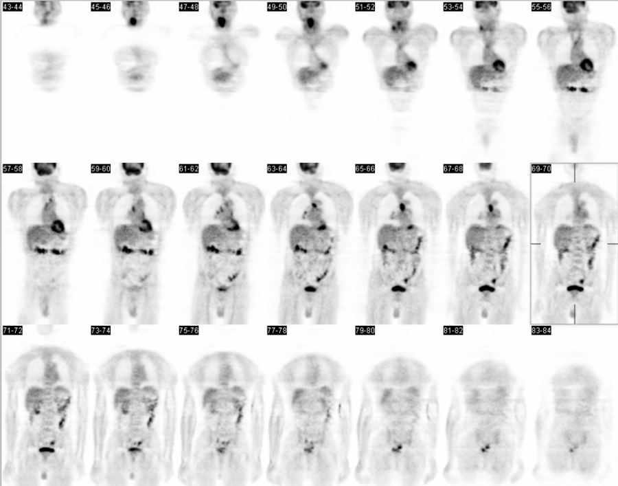
Selected coronal images
View main image(pt) in a separate image viewer
View second image(pt).
Single image from a study prior to PET-CT evaluation
View third image(pt).
Selected axial PET images
View fourth image(pt).
Selected fused PET-CT images
Full history/Diagnosis is available below
Diagnosis: Synchronous primary malignancies of the upper gastrointestinal tract
Full history:
68 year old man with newly diagnosed squamous cell carcinoma of the middle third of the esophagus. A CT examination on 9/8/03 demonstrated a small mediastinal lymph nodes. This examination was obtained for staging.
Radiopharmaceutical:
F-18 FDG i.v.
Findings:
PET-CT:
There is intense metabolic activity associated with the region of circumferential thickening in the mid esophagus at the level of the aortic arch. Multiple metabolically active foci corresponding to lymph nodes are noted in the left pretracheal, right paratracheal, precarinal, right infrahilar and right hilar, and left suprahilar regions.Also, there is a large metabolically active mass lesion identified in the right oropharynx extending to the hypopharynx. There is a focus of increased activity associated with the right scalene muscle, superficial to the metabolically active mass. No definite soft tissue mass is noted in the scalene muscle. Also no metabolically active cervical nodes are noted. No definite metabolically active foci are appreciated in the abdomen or pelvis.On correlation with CT imaging, there are surgical changes identified in the left parietal skull, a pneumatocele is again noted in the superior segment of the left lower lobe. There is an extensive amount of barium retained in the colon from the recent upper GI examination which limits evaluation of the abdomen and pelvis soft tissue structures on the CT examination. A enlarged prostate is appreciated.
Barious Swallow/Upper GI study:
4.5 cm chiefly stricturing esophageal mass at the level of the aortic arch with moderate degree of obstruction.
Discussion:
PET-CT is a well accepted modality for initial staging and restaging evaluation for esophagel carcinoma. Since there is no serosal layer in the esophagus, neoplasm spreads to adjacent tissues and regional lymph nodes early in the course of the disease. (see References below)
Some of the risk factors for esophageal cancer (e.g. smoking) also exist for head and neck tumors such as squamous cell carcinoma.
In this patient, who presented for pre-therapeutic staging for known esophageal cancer, PET-CT evaluation demonstrated a second focus of intense activity in the pharynx, which ultimately turned out to be a second primary malignancy, histologicall different from the known esophageal carcinoma.
Followup:
Pathology report:
1. 09-06-2003 Endoscopic biopsy of the esophagus revealed squamous cell carcinoma, keratinizing, moderately differentiad.
2. 09-16-2003 Laryngoscopic biopsy of the oropharynx revealed oral pharynx, right - Invasive squamous cell carcinoma, poorly differentiated.
3. Bronchospic biopsy of left mainstem bronchus revealed no evidence for malignancy.
Excerpts from patient's discharge summary was as such:
...the two lesions were thought to be two synchronous primary cancers that is both esophageal and the neck cancers were both squamous cell carcinoma not thought to be metastatic from one other...
...the patient was not a candidate for concurrent resections of these primary lesions ...It was then determined that any care at this point would be palliative and not curative.
...The oncology team then agreed to coordinate withradiation oncology the extent of both chemotherapy and radiation therapy for these lesions.
Major teaching point(s):
PET-CT is a very useful modality for the initial and restaging of various neoplasms. Its role in staging esophageal cancers is well established. Pre-treatment PET-CT evaluation can help plan therapy by staging the disease. It can also show additional findings as in this case of two synchronous primary neoplams.
References:
1. Van Westreenen HL, Westerterp M, Bossuyt PM, Pruim J, Sloof GW, Van Lanschot JJ, Groen H, Plukker JT. Systematic Review of the Staging Performance of 18F-Fluorodeoxyglucose Positron Emission Tomography in Esophageal Cancer. J Clin Oncol. 2004 Sep 15;22(18):3805-12.
2. Flamen P, Lerut T, Haustermans K, Van Cutsem E, Mortelmans L. Position of positron emission tomography and other imaging diagnostic modalities in esophageal cancer. Q J Nucl Med Mol Imaging. 2004 Jun;48(2):96-108.
ACR Codes and Keywords:
References and General Discussion of PET Tumor Imaging Studies (Anatomic field:Gasterointestinal System, Category:Neoplasm, Neoplastic-like condition)
Search for similar cases.
Edit this case
Add comments about this case
Return to the Teaching File home page.
Case number: pt117
Copyright by Wash U MO

