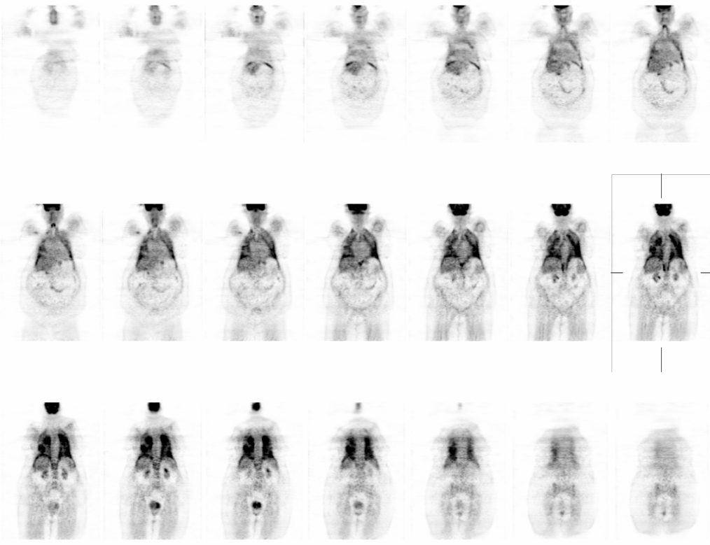

Coronal
View main image(pt) in a separate image viewer
View second image(pt). Axial
View third image(pt). Selected fused PET-CT images
View fourth image(mc). Initial work-up U/S, followed by MRI
Full history/Diagnosis is available below
2. Chest CT (prior to PET: Diffuse patchy, symmetric, ground-glass opacities with upper lobe predominance. A few thickened septal lines are noted, and there are a few small foci of consolidation. These findings are not the expected appearance of lymphangitic spread of tumor. If the patient has been on chemotherapy, these findings can be seen in the setting of acute on chronic hypersensitivity reaction with areas of bronchiolitis obliterans and bronchiolitis obliterans organizing pneumonia. Given the somewhat peripheral distribution in the lung apices, eosinophilic pneumonia may also have a similar appearance.
3. Abdomen ultrasound: Solid spherical mass within the spleen, statistically most likely a benign hemangioma. However, in a person with a personal history of malignancy, evaluation with additional dedicated cross-sectional imaging should be performed.
4. Abdomen MRI: A 6.6 x 5.5 cm heterogeneous lesion demonstrating enhancement in the inferior aspect of the spleen. This most likely represents a benign lesion such as a hamartoma or atypical hemangioma. However, malignancy such as lymphoma and less likely metastatis cannot be excluded. PET imaging may be useful for further evaluation if clinically indicated.
References: 1. Preliminary Findings of a Prospective Study of FDG-PET in Patients with Possible Lung Cancer. Clin Positron Imaging. 2000 Jul;3(4):158.
N.B. The entity known as BOOP is now also referred to as Cryptogenic Organizing Pneumonia.
Radiol Clin North Am. 2001 Nov;39(6):1153-70. High-resolution CT of idiopathic interstitial pneumonias
ORGANIZING PNEUMONIA WITH BOOP-LIKE CHANGES - NO EVIDENCE OF MALIGNANCY
Patient's dyspnea did not resolve even after high dose treatment with prednison. She expired two weeks later.
References and General Discussion of PET Tumor Imaging Studies (Anatomic field:Lung, Mediastinum, and Pleura, Category:Inflammation,Infection)
Return to the Teaching File home page.