Case Author(s): Eric Hutchins, M.D. and Jerold Wallis, M.D.
, 08/25/04 . Rating: #D., #Q.
Diagnosis: Metastatic Melanoma in a Pregnant Patient
Brief history:
23 year-old female with a left lower extremity melanoma removed 12 months ago.
Images:
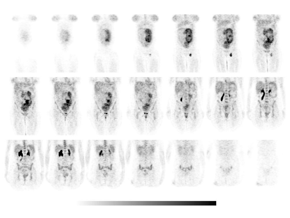
Coronal FDG-PET images.
View main image(pt) in a separate image viewer
View second image(pt).
Coronal FDG-PET images of the lower extremities.
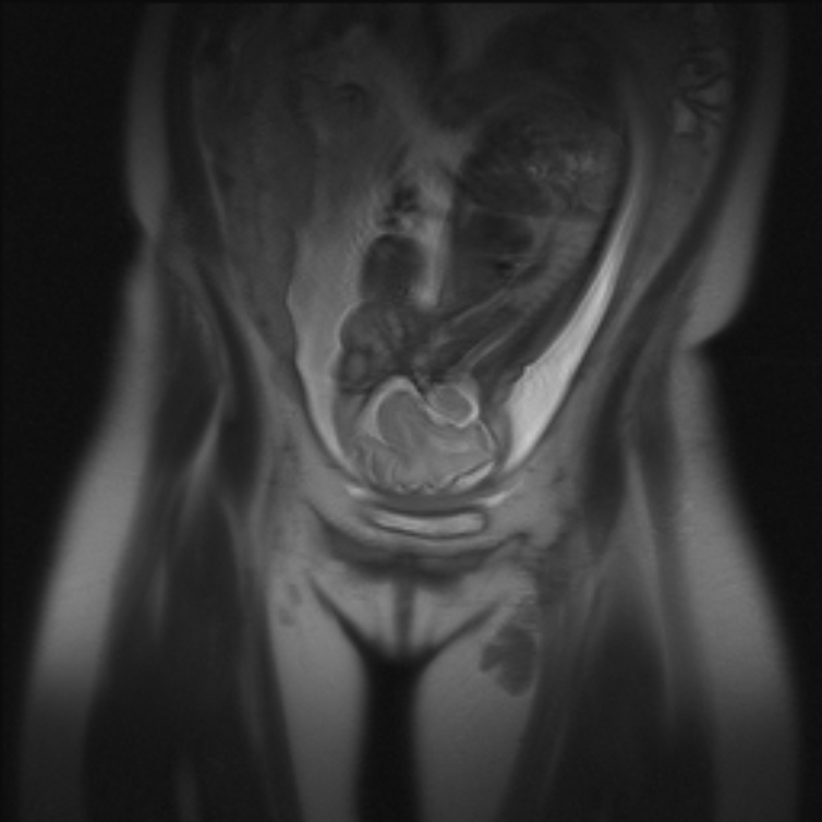
View third image(mr).
Coronal T2 weighted MR image.
Full history/Diagnosis is available below
Diagnosis: Metastatic Melanoma in a Pregnant Patient
Full history:
23 year-old female a left lower extremity melanoma removed 12 months ago. Her clinical findings include a healing skin graft on the medial aspect of her left calf and a new palpable mass in the left groin.
Radiopharmaceutical:
F-18 fluorodeoxyglucose i.v.
Findings:
Intense hypermetabolism within a group of four enlarged left inguinal lymph nodes is seen on both sets of coronal images suggesting nodal metastasis. There is no evidence of other metastatic disease. FDG uptake is also seen in the placenta and fetus of this patient who is seven months pregnant. The coronal PET images of the legs also demonstrate mild hypermetabolism in the soft tissues of the medial left calf at the site of the primary tumor resection. This is considered to be post-surgical healing. The coronal T2 weighted MR image demonstrates the fetus as well as the left inguinal lymphadenopathy.
Discussion:
FDG-PET imaging can be helpful in detecting locoregional disease and distant metastases in patients with known melanoma and clinical suspicion of tumor recurrence. In a recent study of 156 patients, FDG-PET was more accurate (91% vs. 67%) than standard diagnostic procedures (physical examination, radiography, CT, bone scanning, MRI, ultrasound, and hepatic enzyme assays) at detecting metastases. Lung nodules are an exception , as CT is more sensitive than FDG-PET (93% vs. 57%). Also, in the initial staging of melanoma, a sentinel lymph node procedure can detect micrometastases to local nodes much better than FDG-PET.
FDG-PET imaging can be performed in pregnant patients if necessary. The fetal radiation dose from FDG at 6-9 months gestation is 0.017 mGy/MBq (about 60 mrem/mCi), and is about 30% higher in early pregnancy. Useful methods of minimizing the dose to the fetus include using PET rather than PET/CT, using only 8-10 mCi, placing a foley catheter to drain radioactive urine, and using intravenous fluids to help clear activity quickly.
References:
Fuster D, Chiang S, Johnson G, Schuchter LM, Zhuang H, Alavi A. Is 18F-FDG PET more accurate than standard diagnostic procedures in the detection of suspected recurrent melanoma? J Nucl Med. 2004 Aug;45(8):1323-7
Stabin, Michael G. Proposed Addendum to Previously Published Fetal Dose Estimate Tables for 18F-FDG. J Nucl Med. 2004 Vol. 45 No. 4 634-635
ACR Codes and Keywords:
References and General Discussion of PET Tumor Imaging Studies (Anatomic field:Vascular and Lymphatic Systems, Category:Misc)
Search for similar cases.
Edit this case
Add comments about this case
Return to the Teaching File home page.
Case number: pt114
Copyright by Wash U MO