Case Author(s): Eric Hutchins, M.D. and Farrokh Dehdashti, M.D. M.D. , 08/19/04 . Rating: #D., #Q.
Diagnosis: Mycobacterium Avium Complex
Brief history:
75-year-old female with a solitary pulmonary nodule.
Images:
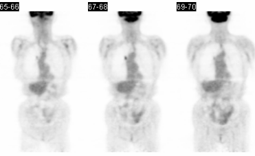
Coronal images from FDG-PET scan.
View main image(pt) in a separate image viewer
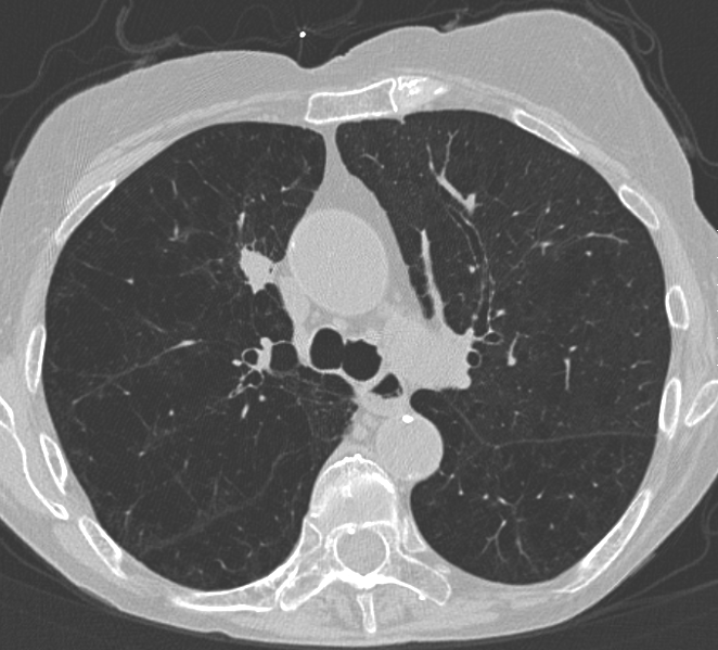
View second image(ct).
Initial CT images at time of PET imaging.
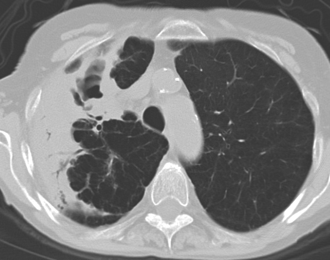
View third image(ct).
CT performed 3 months after PET imaging.
Full history/Diagnosis is available below
Diagnosis: Mycobacterium Avium Complex
Full history:
75-year-old female with COPD initially presented with dyspnea. A chest radiograph demonstrated a new right upper lobe nodule compared with a previous chest radiograph performed 3 years prior. A CT of the thorax showed a noncalcified 1.1 x 1.4 cm nodule in the right upper lobe and underlying emphysematous lung disease. FDG-PET images demonstrated increased metabolism (SUVmax = 3.8) within the nodule suggesting malignancy. Patient refused surgery and returned 3 months later with complaints of dyspnea and weight loss.
Radiopharmaceutical:
14.3 mCi F-18 Fluorodeoxyglucose I.V.
Findings:
PET images show increased metabolism (SUVmax = 3.8) within the right upper lobe pulmonary nodule. Initial CT images show a noncalcified 1.1 x 1.4 cm nodule in the right upper lobe corresponding to the increased metabolism seen on PET imaging. Also noted are diffuse emphysematous changes of the lungs. Three months later, a CT of the chest shows interval development of a right apical cavitary lesion with surrounding areas of airspace disease as well as enhancement and pleural thickening. The previously seen right upper lobe cavitary lesion has filled with fluid. Such quick progression of disease favors an inflammatory process over malignancy. Subsequently, the patient underwent bronchoscopy and biopsy demonstrated granulomatous inflammation containing acid-fast bacilli. Mycobacterium avium complex was eventually identified by the Missouri Tuberculosis Reference Lab.
Discussion:
Pulmonary nontuberculous mycobacterial infections can have many radiographic manifestations. The classic infection is an elderly white man with underlying lung disease, such as COPD. "The most common findings are heterogeneous linear and nodular areas of increased opacity in the apical and posterior segments of the upper lobes with or without calcification." Cavitation is common and leads to endobronchial spread of mycobacteria, which causes scattered nodules in a centrilobular distribution.
While an SUV of less than 2.5 is quite sensitive for benign pulmonary lesions, active inflammatory disorders such as tuberculosis, histoplasmosis, aspergillosis, other infections,and eosinophilic lung disease can demonstrate significant hypermetabolism. Lesions with an SUV of greater than 2.5 are "considered malignant until proven otherwise." This patient is an example of a false-positive study when evaluating a solitary pulmonary nodule for malignancy.
References
Erasmus JJ, McAdams HP, Farrell MA: Pulmonary nontuberculous mycobacterial infection. Radiographics 1999; 19(6): 1487-505.
Patz EF,Lowe JM, Hoffmann JM, et al. Focal pulmonary abnormalities: evaluation with F-18 fluorodeoxyglucose PET scanning. Radiology 1993;188:487-490.
Differential Diagnosis List
Lung carcinoma, tuberculosis and NTMB infection, fungal infection (especially histoplasmosis and aspergillosis), other infections
ACR Codes and Keywords:
References and General Discussion of PET Tumor Imaging Studies (Anatomic field:Lung, Mediastinum, and Pleura, Category:Inflammation,Infection)
Search for similar cases.
Edit this case
Add comments about this case
Return to the Teaching File home page.
Case number: pt113
Copyright by Wash U MO