Case Author(s): Randal Anderson, M.D. and Farrokh Dehdashti, M.D. , 10/14/03 . Rating: #D3, #Q4
Diagnosis: Metastatic cervical cancer.
Brief history:
54 year old woman with newly discovered cancer. She has not yet received therapy. We are asked to evaluate extent of disease.
Images:
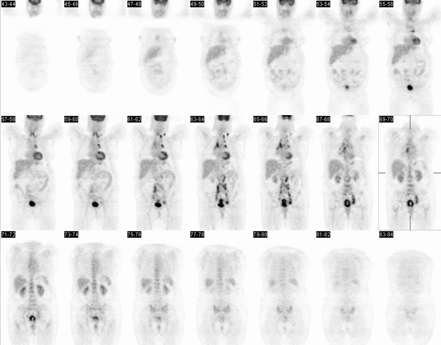
Coronal PET images
View main image(pt) in a separate image viewer
View second image(pt).
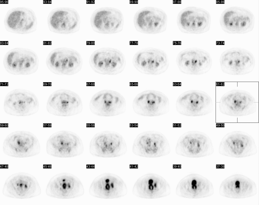
Axial PET images
View third image(ct).
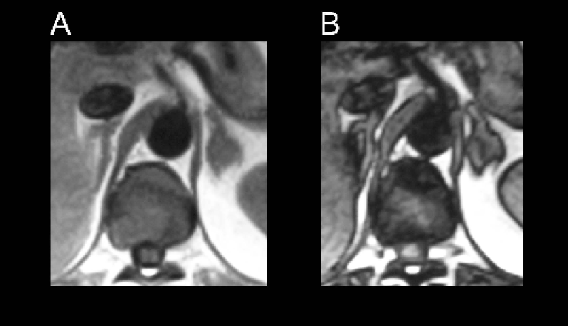
Axial CT slice through the pelvis
View fourth image(pt).
PET-CT fusion images
Full history/Diagnosis is available below
Diagnosis: Metastatic cervical cancer.
Full history:
54 year old woman with newly discovered cervical cancer. She has not yet received therapy. We are asked to evaluate extent of disease.
Radiopharmaceutical:
F-18 fluorodeoxyglucose (FDG)
Findings:
PET images demonstrated Large soft tissue mass with increased activity in the region of the cervix, consistent with the patient's known cervical cancer. In addition, increased uptake is seen in bilateral obturator nodes, left external iliac nodes, bilateral common iliac nodes, bilateral paraaortic nodes (greater on the left than the right), and a probable celiac node. There is also a presacral soft tissue nodule at the level of the sacral promontory just to the right of midline with increased activity, also likely metastatic disease. In the chest, lymph nodes with increased activity are seen in the left supraclavicular region (at least two nodes), right paratracheal region, prevascular region, precarinal region, and subcarinal region.
CT of the chest, abdomen and pelvis demonstrated extensive bulky retroperitoneal and pelvic lymphadenopathy. This lymphadenopathy is seen in the periaortic, aortocaval, iliac, and obturator chain. The lymph nodes in the chest were not identified as abnormal on the CT scan; although, in retrospect they were abnormal.
Discussion:
Lymph node involvement is very common in cervical cancer and the extent of lymph node involvement on FDG-PET has been shown to be predictive of prognosis. FDG-PET has been shown to be superior to CT is identifying lymph node metastasis and predicting prognosis.
Grigsby PW, Siegel BA, Dehdashti F. Lymph node staging by positron emission tomography in patients with carcinoma of the cervix. J Clin Oncol 2001; 19:3745-3749.
Tran B, Grigsby PW, Dehdashti F, Herzog T, Siegel BA. Occult supraclavicular lymph node metastasis identified by FDG-PET in patients with carcinoma of the uterine cervix. Gynecol Oncol 2003; 90:572-576.
Followup:
Biopsy of cervical mass showed poorly differentiated carcinoma. Patient passed way a few months after diagnosis.
Major teaching point(s):
Demonstration of extensive lymph node involvement in a pattern typical for cervical cancer.
ACR Codes and Keywords:
References and General Discussion of PET Tumor Imaging Studies (Anatomic field:Genitourinary System, Category:Neoplasm, Neoplastic-like condition)
Search for similar cases.
Edit this case
Add comments about this case
Return to the Teaching File home page.
Case number: pt107
Copyright by Wash U MO