Case Author(s): Feiyu Xue, M.D., Ph.D., Barry Siegel, MD, and Tom Miller, M.D., Ph.D. , . Rating: #D2, #Q3
Diagnosis: Atypical Mycobacterial (MAI) Infection
Brief history:
67 year-old woman with a history of ovarian cancer presents for evaluation of multiple, newly discovered pulmonary nodules.
Images:
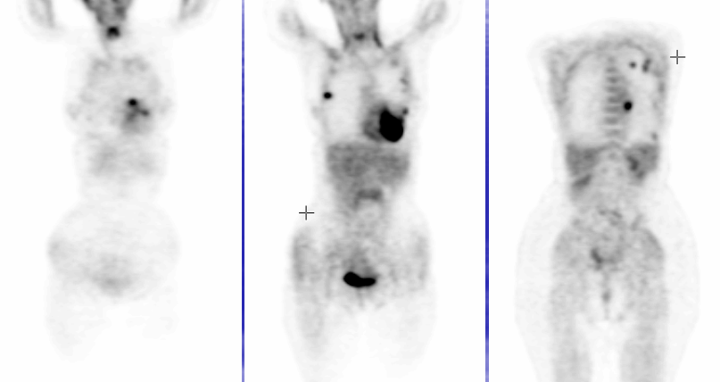
FDG-PET Coronal images
View main image(pt) in a separate image viewer
View second image(pt).
FDG-PET axial images
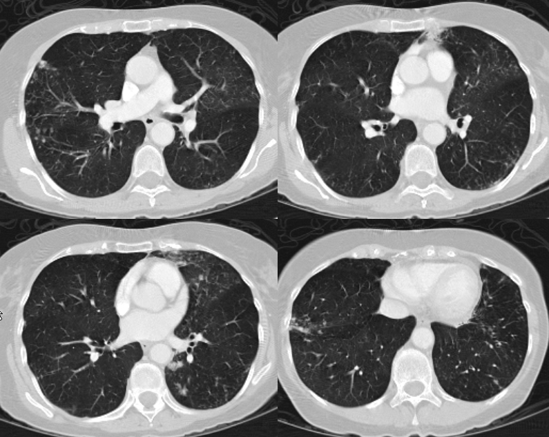
View third image(ct).
Lung windows of CT of the chest
Full history/Diagnosis is available below
Diagnosis: Atypical Mycobacterial (MAI) Infection
Full history:
A 67 year old woman has a history of ovarian cancer, treated 5 years ago by total abdominal hysterectomy, bilateral salpingo-oophorectomy, and chemotherapy. She was found to have multiple bilateral pulmonary nodules/opacities on a recent surveillance CT study. FDG-PET was requested for further evaluation.
Radiopharmaceutical:
F-18 FDG
Findings:
There are multiple foci of increased FDG uptake in the lungs bilaterally. CT shows multiple nodular infiltrates in the lungs bilaterally, involving the lingula and superior segment of the left upper lobe, right upper lobe and right middle lobe, correlating with the foci of increased activity seen on FDG-PET. In addition, there are scattered ground-glass opacities and nodular infiltrates. Some areas of nodular infiltrate demonstrate a “tree-in-bud” appearance.
Discussion:
In this patient with a remote history of cancer, metastatic disease has to be considered. However, pulmonary metastasis without either nodal disease in the pelvis or retroperitoneum or peritoneal implants would be highly unusual in ovarian cancer. Infections, including tuberculosis, atypical mycobacterial infections, and fungal infection, can also present with an identical appearance on PET and CT. The finding of ground-glass opacities suggests an acute process. Nodular infiltrates with a “tree-in-bud” appearance, especially in the cardiophrenic angle of the lingula, although not specific, are quite typical of Mycobacteriun avium intracellulare (MAI) infection. The fact that this patient is a relatively asymptomatic elderly woman is also suggestive of an atypical mycobacterial infection.
MAI infection causes two patterns of pulmonary disease in two distinct populations of patients. The fibrocavitary pattern with radiologic similarities to tuberculosis is seen in patients with a history of tobacco and/or alcohol abuse. Nodular infiltrates as seen in this case are the typical appearance primarily seen in elderly women without an obvious immunodeficiency disorder.
Followup:
This patient underwent bronchoscopy and bronchial lavage. MAI infection was confirmed by culture.
Major teaching point(s):
The clues to the correct diagnosis in this case are found in the clinical history and correlative CT findings. It is unusual for ovarian cancer to metastasize to the lungs without nodal disease or widespread peritoneal metastases. CT of the chest showed characteristic (but non-specific) findings of ground-glass opacities and nodular infiltrates with a “tree-in-bud” appearance. The CT appearance, although non-specific, is quite typical of mycobacterial infection.
References
1. Bandoh S, Fujita J, Ueda Y, et al. Uptake of fluorine-18-fluorodeoxyglucose in pulmonary Mycobacterium avium complex infection. Intern Med 2003; 42:726-9.
2. Bleeker-Rovers CP, de Kleijn EM, Corstens FH, van der Meer JW, Oyen WJ. Clinical value of FDG PET in patients with fever of unknown origin and patients suspected of focal infection or inflammation. Eur J Nucl Med Mol Imaging 2004; 31:29-37.
3. Jeong YJ, Lee KS, Koh WJ, Han J, Kim TS, Kwon OJ. Nontuberculous mycobacterial pulmonary infection in immunocompetent patients: comparison of thin-section CT and histopathologic findings. Radiology 2004; 231:880-6.
4. Prince, DS, Peterson, DD, Steiner, RM, et al Infection with mycobacterium avium complex in patients without predisposing conditions. N Engl J Med 1989; 321,863-8.
5. Yang CM, Hsu CH, Lee CM, Wang FC. Intense uptake of
Differential Diagnosis List
Neoplasm (metastasis)
Infection (TB, MAI, fungal)
Inflammation (sarcoidosis, silicosis, Wegener’s granulomatosis)
ACR Codes and Keywords:
References and General Discussion of PET Tumor Imaging Studies (Anatomic field:Lung, Mediastinum, and Pleura, Category:Inflammation,Infection)
Search for similar cases.
Edit this case
Add comments about this case
Return to the Teaching File home page.
Case number: pt105
Copyright by Wash U MO