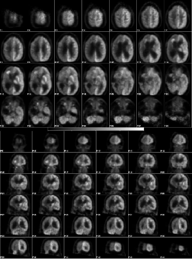
Axial and coronal FDG-PET brain images
View main image(pt) in a separate image viewer
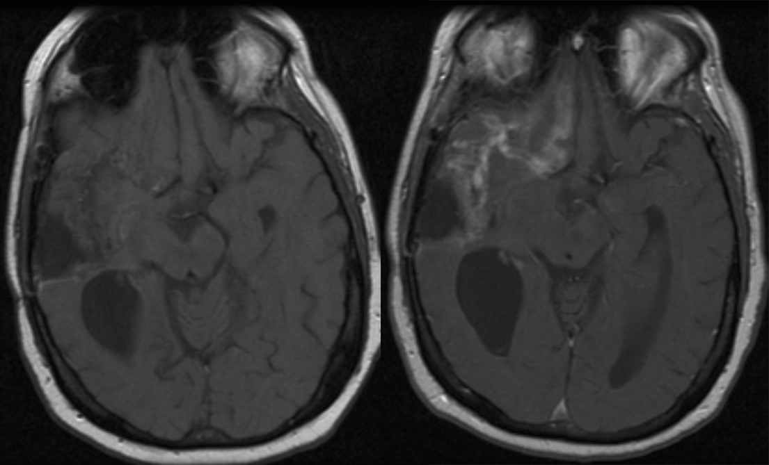
View second image(mr). Selected T1-weighted pre -and post-gadolinum axial MR images of the brain.
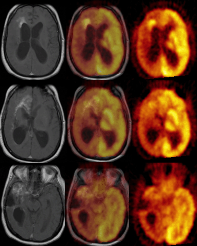
View third image(pt). Selected T1-weighted post-gadolinum axial MR images with corresponding fused and PET axial images
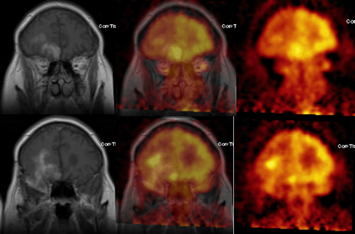
View fourth image(pt). Selected T1-weighted post-gadolinum coronal MR images with corresponding fused and PET coronal images
Full history/Diagnosis is available below
PET: There is generalized mild decrease in FDG uptake in the right cerebral hemisphere suggesting edema and/or post-radiation effect. There is decreased FDG uptake in the contralateral left cerebellar hemisphere, consistent with crossed cerebellar diaschisis. There is increased FDG uptake corresponding to the abnormal enhancing masses in the right anterior and inferior frontal lobe, anterior to the frontal horn of the lateral ventricle and anterior to the right temporal lobectomy surgical bed. There is enlargement of the lateral and third ventricle consistent with hydrocephalus.
FDG-PET can also assist in defining the most metabolically active sites for stereotactic biopsy. Coregistration of the MRI and FDG-PET images is essential for accurate evaluation and localization of brain tumors.
FDG PET has also been demonstrated to be useful in distinguishing between recurrent high-grade tumor and radiation necrosis. In this case, there is relatively low background cortical activity in the right frontotemporal region, as a result of edema or post-radiation effect. This facilitates detection of recurrent high grade tumor.
With low-grade brain tumors, FDG-PET has limited value in defining extent of tumor involvement and recurrence.
Positron-emitting amino acids, such as C-11 methionine, have demonstrated accumulation in brain tumors and have the advantage of low background cortical activity. The relationship between degree of C-11 methionine uptake and tumor grade is not established.
REFERENCES
Benard F, Romsa J, Hustinx R. Imaging gliomas with positron emission tomography and single-photon emission computed tomography. Semin Nucl Med. 2003; 33:148-62.
Wong TZ, van der Westhuizen GJ, Coleman RE. Positron emission tomography imaging of brain tumors. Neuroimaging Clin N Am. 2002; 12:615-26
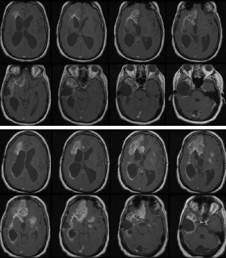
View followup image(mr). Selected T1-weighted post-gadolinum axial MR images. Top 8 images: Obtained concominantly as the above PET examination. Bottom 8 images: Obtained 2 months after the PET examination.
2. Coregistration of MR and FDG-PET images is essential for accurate evaluation and localization of brain tumors.
References and General Discussion of PET Tumor Imaging Studies (Anatomic field:Skull and Contents, Category:Neoplasm, Neoplastic-like condition)
Return to the Teaching File home page.