Case Author(s): Jayson R. St. Jacques, M.D. and Jerold Wallis, M.D. , 6/6/03 . Rating: #D2, #Q5
Diagnosis: Recurrent Sigmoid Colon Cancer with Small Bowel Obstruction
Brief history:
75 year old woman with history of sigmoid colon cancer, post partial colectomy and chemotherapy 5 years ago.
Images:

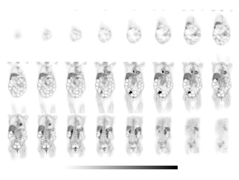
Coronal FDG-PET images.
View main image(pt) in a separate image viewer
View second image(xr).
AP Abdominal Radiograph
View third image(mr).
Coronal MR image.
Full history/Diagnosis is available below
Diagnosis: Recurrent Sigmoid Colon Cancer with Small Bowel Obstruction
Full history:
75 year old woman with history of sigmoid colon adenocarcinoma. She underwent partial colectomy and chemotherapy 5 years ago and now presents with abdominal pain.
PET imaging was requested to evaluate for recurrent disease.
Radiopharmaceutical:
15.0 mCi of Fluorodeoxyglucose i.v.
Findings:
Focal uptake within the pelvis consistent with recurrence. Multiple loops of dilated bowel consistent with high grade obstruction. Focal uptake in the region of the right heart of uncertain significance, but may represent a metastasis.
Followup:
The patient underwent surgery to remove the focus of recurrence and to relieve the bowel obstruction.
The patient also underwent CT and MR imaging to evaluate the abnormal focus of increased uptake in the region of the right heart. There was no CT correlate for this finding and the MR had to be terminated early secondary to patient discomfort.
Follow up cardiac perfusion imaging demonstrated mild distal septal and apical ischemia (link below).
View followup image(mi).
3D display of myocardial SPECT rest/stress images with thallium/sestamibi dual isotope imaging.
Major teaching point(s):
The appearance of bowel obstruction on PET imaging is not a common finding, but is quite distinctive when present. PET imaging is useful for restaging colon cancer.
ACR Codes and Keywords:
References and General Discussion of PET Tumor Imaging Studies (Anatomic field:Gasterointestinal System, Category:Neoplasm, Neoplastic-like condition)
Search for similar cases.
Edit this case
Add comments about this case
Return to the Teaching File home page.
Case number: pt100
Copyright by Wash U MO

