Case Author(s): Brock G. McDaniel, M.D. and Tom R. Miller, M.D., Ph.D. , . Rating: #D3, #Q3
Diagnosis: Stage IIB cervical carcinoma with concomitant pneumonia
Brief history:
61-year-old women with stage IIB cervical carcinoma diagnosed in 2001.
Images:
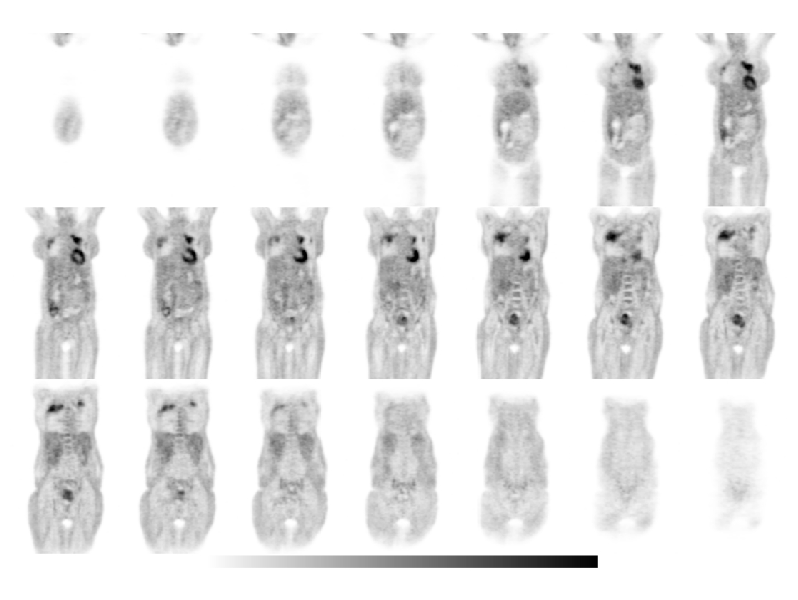
Coronal PET (03/31/03)
View main image(pt) in a separate image viewer
View second image(ct).
Pelvic CT (03/26/03)
View third image(xr).
CXR (03/25/03)
View fourth image(xr).
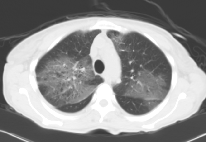
Chest CT (04/02/03)
Full history/Diagnosis is available below
Diagnosis: Stage IIB cervical carcinoma with concomitant pneumonia
Full history:
61-year-old women with stage IIB cervical carcinoma diagnosed in 2001 who originally declined therapy. She now presents with vaginal bleeding and requests treatment. She has an elevated white blood cell count (WBC=19.2k with 92% neutrophils).
Radiopharmaceutical:
15.0 mCi F-18 Fluorodeoxyglucose i.v.
Findings:
The PET images demonstrate multifocal hypermetabolism within the lung apices bilaterally. A plain radiograph of the chest performed the day of the PET scan (not shown), shows bi-apical patchy opacities which were not seen on a previous chest radiograph (1 week earlier). A follow-up chest CT demonstrated bilateral upper lobe ground glass opacities consistent with an acute infectious/inflammatory process.
Within the pelvis is a heterogenous focus of increased FDG uptake consistent with the known cervical carcinoma. CT images confirm the cervical mass.
Discussion:
The utility of PET in the diagnoses and staging of cervical cancer is well known. However, increased uptake of F18-Fluorodeoxyglucose is not specific for cancer. Many acute or chronic inflammatory processes where there are an increased number of neutrophils or activated macrophages will also demonstrate increased uptake on FDG-PET. Although malignant involvement can not be completely excluded, an important clue that this uptake represents an acute inflammatory process is the rapid appearance of patchy opacities on chest radiographs. Such a rapid progression would not be expected with metastasis.
Followup:
Given the appearance of these bi-apical opacities, the rapid development, the elevated white blood cell count and the patient's potential to be in an immunocompromised state, serial sputum samples were sent for acid fast staining analysis to exclude tuberculosis. All samples failed to grow anything and no acid fast positive bacilli were seen. The patient began to improve on empirical antibiotic therapy (Rifampin, Isoniazide, Pyraxinamide) and her leukocytosis began to resolve. The follow-up chest radiographs demonstrated interval improvement in her opacities.
Differential Diagnosis List
DDx: Atypical pneumonia, tuberculosis or other infectious or inflammatory process with pulmonary/pleural metastasis and primary lung cancer less likely. In the right clinical setting radiation pneumonitis, drug reaction, or talc pleurodesis could have this appearance.
ACR Codes and Keywords:
References and General Discussion of PET Tumor Imaging Studies (Anatomic field:Genitourinary System, Category:Neoplasm, Neoplastic-like condition)
Search for similar cases.
Edit this case
Add comments about this case
Return to the Teaching File home page.
Case number: pt099
Copyright by Wash U MO